DIGITAL ORTHO LAB
Boneborne Rapid Palatal Expansion—the Virtual Way
Digitalization is changing not only our society in general, but dentistry in particular. In orthodontics, although one might have the impression that digitalization refers exclusively to aligners—since they occupy a dominant position in the media—the spectrum of digital orthodontics is almost unlimited. Computer-aided design and manufacturing (CAD/CAM) is facilitating a high level of innovation in orthodontics by optimizing proven diagnostic and therapeutic procedures.
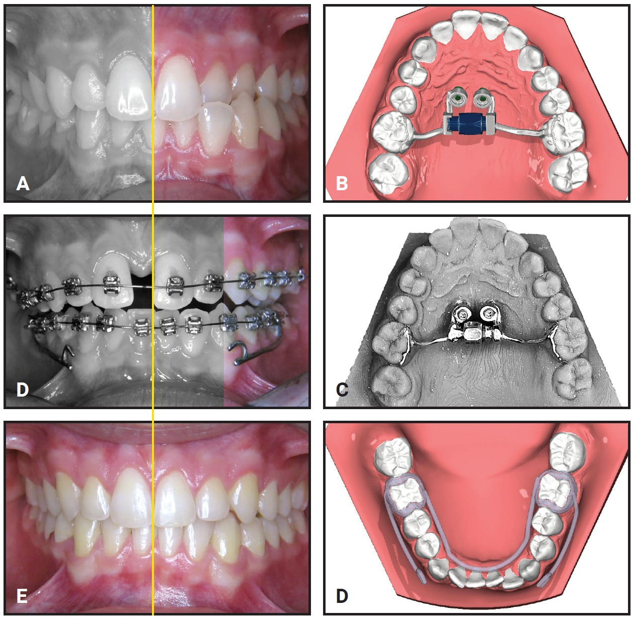
In this first edition of the Digital Ortho Lab, we will describe the procedure for virtually designing a boneborne rapid palatal expander, often called a mini-implant-assisted rapid palatal expander (MARPE). The technique is illustrated by selected diagnostic records of a 15-year-old male patient who presented with a unilateral crossbite and a Class III tendency.
Why MARPE?
Conventional rapid palatal expansion produces such side effects as tipping of teeth, a reduction in buccal bone thickness, and a loss of marginal bone height resulting in gingival recession.1-4 Boneborne expanders offer several advantages, including more predictable skeletal expansion and fewer dental side effects. MARPE, which seems to be especially well suited for patients during the postpubertal growth spurt,5 has been associated with a high success rate.6
For many years, MARPE appliances were prefabricated products. The expansion appliance was usually inserted first, followed by two to four temporary anchorage devices (TADs)—as with the maxillary skeletal expander (MSE).7 In the digital world, however, the appliance is always fully customized for the individual patient (Fig. 1).
Appliance Design
To virtually plan and design a boneborne expander, the digital diagnostic records need to be matched (superimposed) using suitable software (Fig. 2). Recommended records are an intraoral scan and corresponding photos, as well as a lateral cephalogram and, if indicated, cone-beam computed tomography (CBCT).
Similar articles from the archive:
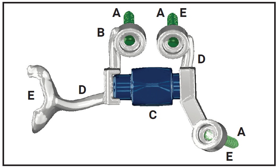
Fig. 1 Basic components of mini-implant-supported rapid palatal expander (MARPE). A. Temporary anchorage devices (TADs). B. Abutments. C. Expansion screw.** D. Connecting elements. E. Posterior anchorage (anti-tipping) from molar pads or TADs.
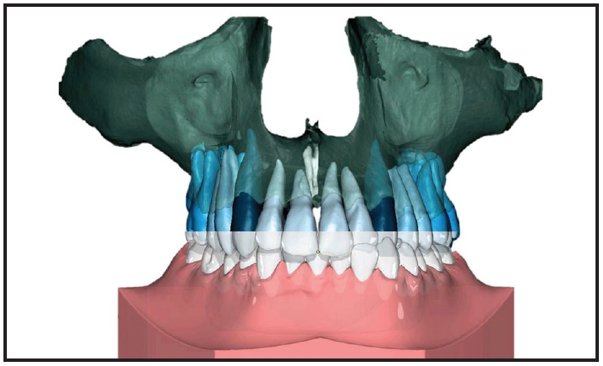
Fig. 2 Initial intraoral scan of 15-year-old male patient with unilateral crossbite and Class III tendency matched with cone-beam computed tomography.
There are many software products on the orthodontic market suitable for designing any kind of MARPE (Table 1); OnyxCeph3‡ was utilized for this patient. Pure CAD suites can support the more advanced operator by facilitating individual functions such as finite-element analysis.
In the first step of appliance planning, the “virtual TADs” are digitally positioned (Fig. 3). The virtual TADs should be surrounded by adequate bone while avoiding critical anatomical structures such as teeth and nerves and respecting the soft tissue. Bicortical positioning is often helpful.8 The TAD length, diameter, and design must be adapted to the individual patient’s anatomy9 since, for example, the vertical bone height is often reduced in the posterior palate.10 The insertion angle seems to be crucial. TAD placement can be optimized and adapted to varying bone thicknesses if the bone morphology of the palate is properly evaluated (preferably with CBCT).11,12
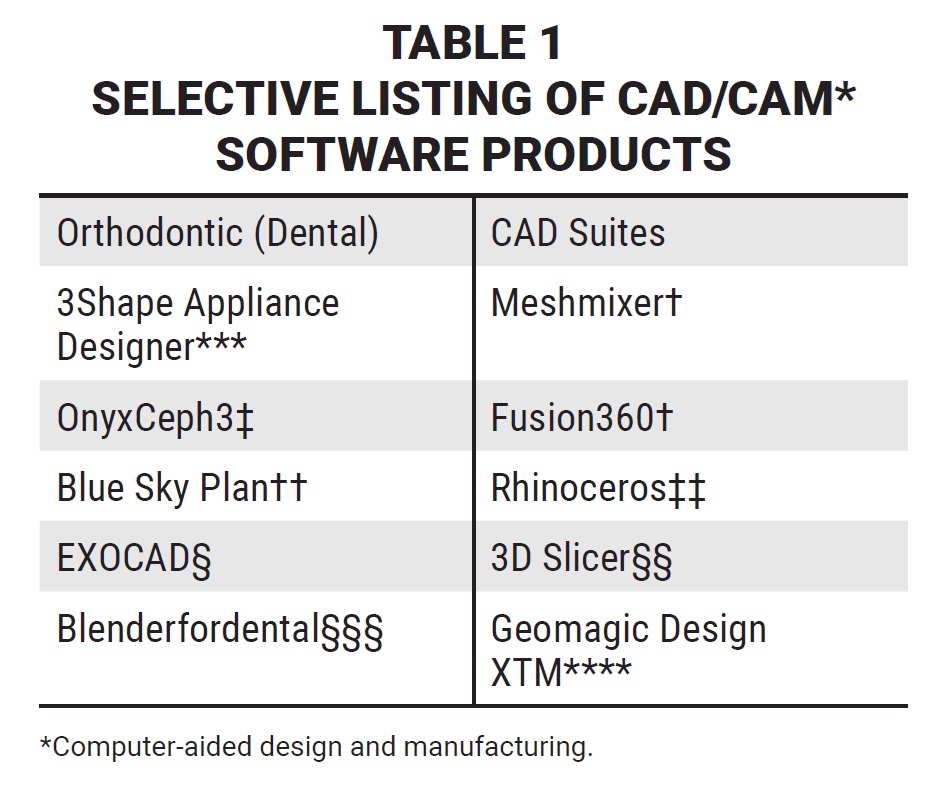
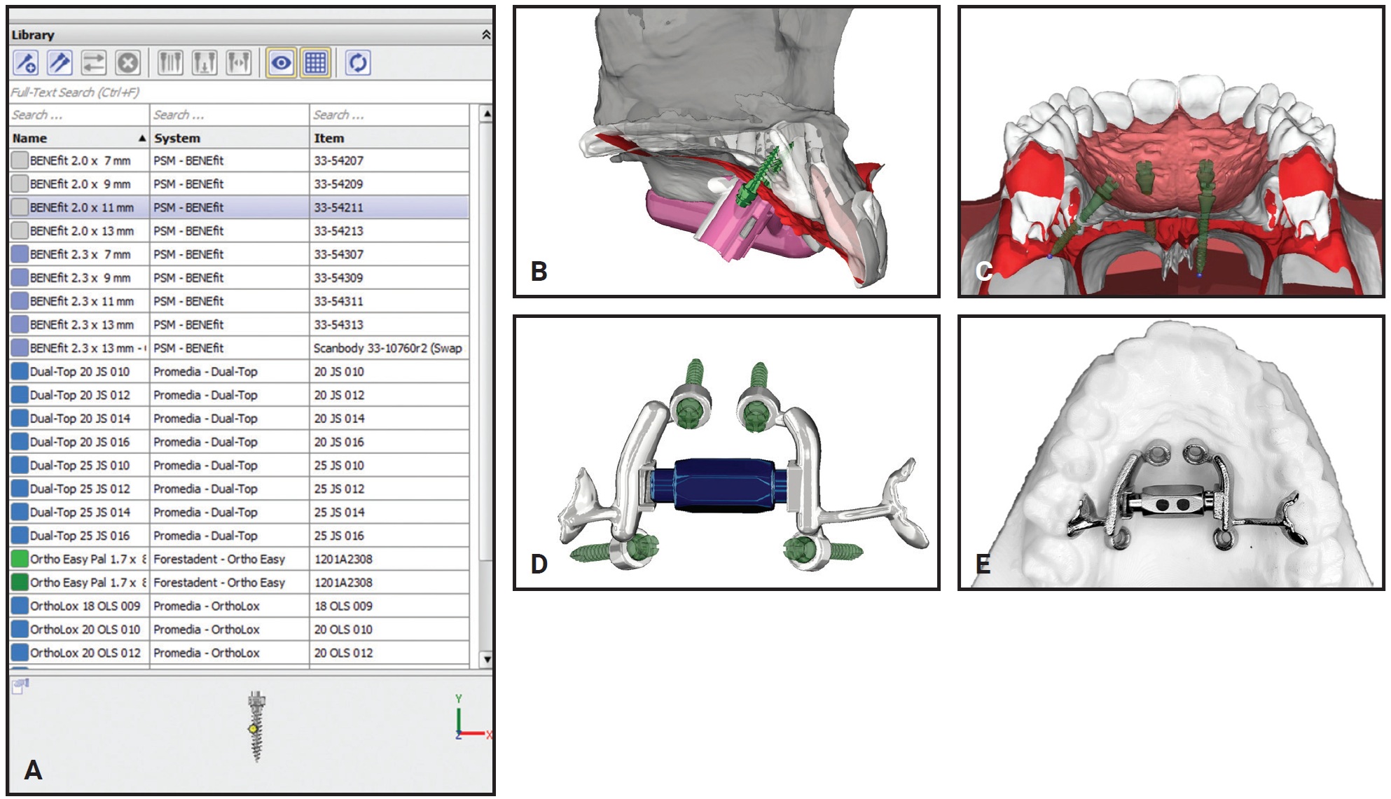
Fig. 3 “Virtual TAD” design. A. Software library includes most commercially available TADs. B. TADs virtually positioned and digital insertion guide simultaneously designed for chairside insertion. C. Left side: posterior screws too long for reduced posterior bone; right side: screw positions customized with respect to hard and soft tissues. D. Power Expander14 MARPE serves as own insertion guide. E. Power Expander three-dimensionally printed by selective laser melting (SLM).
Once the virtual TAD location is established, a digital insertion guide is designed for three-dimensional printing, either in-house or in a laboratory. The guide allows the MARPE and miniscrews to be installed in one session,1,2,8,9 which requires careful digital planning for anatomical site transfer.10,13 This aspect will be covered in more detail in future Digital Ortho Lab articles.
The MARPE is then digitally constructed with respect to the selected mini-implant positions. Many designs have been developed over the past two decades: Wilmes and colleagues introduced the Hybrid Hyrax§§§§ expander in 2007,15,16 and similar hybrid expanders were published by Garib and colleagues in 2008,17 Lee and colleagues in 2010,18 Winsauer and colleagues in 2013,19 and Moon and colleagues in 2015 (the MSE).20 Interestingly, because of their fully customized, patient-centered 3D design, the MARPE appliances have gradually become more difficult to differentiate. Download the STL file for a Hybrid Hyrax§§§§ MARPE15,16 and watch the video below for an example of creating a design using OnyxCeph3‡ software.
Finally, the STL files for the MARPE are sent to a 3D metal-printing service that uses selective laser melting.1,2 Metal printing enables infinitely customizable CAD/CAM designs to be produced for clinical usage.
The 15-year-old patient shown in Figure 2 was treated with a Hybrid Hyrax and Class III elastics, utilizing a CAD/CAM-produced Tandem Appliance (Fig. 4).21 Keep in mind, however, that while the digital lab clearly optimizes diagnostic and therapeutic procedures, the technology does not perform the treatment. Only the orthodontic specialist can do that, with the support of CAD/CAM tools and relevant scientific findings.
FOOTNOTES
- **PowerScrew, Tiger Dental, Bregenz, Germany; www.tigerdental.com.
- ***Trademark of 3Shape, Copenhagen, Denmark; www.3shape.com.
- †Autodesk, Inc., San Rafael, CA; www.autodesk.com.
- ‡OnyxCeph, Chemnitz, Germany; www.onyxceph.de.
- ††Blue Sky Bio, Libertyville, IL; www.blueskyplan.com/orthodontic.
- ‡‡Robert McNeel & Associates, Seattle, WA; www.rhino3d.com.
- §Registered trademark of Align Technology, Inc., San Jose, CA; www.aligntech.com.
- §§3D Slicer is a free, open-source, multi-platform software package; www.slicer.org.
- §§§Blender for Dental International, Brisbane, Australia; www.blenderfordentala.com.
- ****Registered trademark of 3D Systems, Inc., Rock Hill, SC; www.3Dsystems.com.
- §§§§Registered trademark of Dentaurum, Inc., Newtown, PA; www.dentaurum.com.
- †††SLM 50, Realizer, DW Lingual Systems GmbH, Bad Essen, Germany; www.lingualsystems.de.
REFERENCES
- 1. Garib, D.G.; Henriques, J.F.C.; Janson, G.; Freitas, M.R.; and Fernandes, A.Y.: Periodontal effects of rapid maxillary expansion with tooth-tissue-borne and tooth-borne expanders: A computed tomography evaluation, Am. J. Orthod. 129:749-758, 2006.
- 2. Schuster, G.; Borel-Scherf, I.; and Schopf, P.M.: Frequency of and complications in the use of RPE appliances—results of a survey in the Federal State of Hesse, Germany, J. Orofac. Orthop. 66:148-161, 2005.
- 3. Erverdi, N.; Tosun, T.; and Keles, A.: A new anchorage site for the treatment of anterior open bite: Zygomatic anchorage, World J. Orthod. 3:147-153, 2002.
- 4. Akyalcin, S.; Schaefer, J.S.; English, J.D.; Stephens, C.R.; and Winkelmann, S.: A cone-beam computed tomography evaluation of buccal bone thickness following maxillary expansion, Imaging Sci. Dent. 43:85-90, 2013.
- 5. Jia, H.; Zhuang, L.; Zhang, N.; Bian, Y.; and Li, S.: Comparison of skeletal maxillary transverse deficiency treated by microimplant-assisted rapid palatal expansion and tooth-borne expansion during the post-pubertal growth spurt stage, Angle Orthod. 91:36-45, 2021.
- 6. Kapetanović, A.; Theodorou, C.I.; Bergé, S.J.; Schols, J.G.J.H.; and Xi, T.: Efficacy of miniscrew-assisted rapid palatal expansion (MARPE) in late adolescents and adults: A systematic review and meta-analysis, Eur. J. Orthod. 43:313-323, 2021.
- 7. Carlson, C.; Sung, J.; McComb, R.W.; Machado, A.W.; and Moon, W.: Microimplant-assisted rapid palatal expansion appliance to orthopedically correct transverse maxillary deficiency in an adult, Am. J. Orthod. 149:716-728, 2016.
- 8. Lee, R.J.; Moon, W.; and Hong, C.: Effects of monocortical and bicortical mini-implant anchorage on bone-borne palatal expansion using finite element analysis, Am. J. Orthod. 151:887-897, 2017.
- 9. Nojima, L.I.; Nojima, M.C.G.; Carneiro da Cunha, A.; Guss, N.O.; and Sant’Anna, E.F.: Mini-implant selection protocol applied to MARPE, Dent. Press J. Orthod. 23:93-101, 2018.
- 10. Kang, S.; Lee, S.J.; Ahn, S.J.; Heo, M.S.; and Kim, T.W.: Bone thickness of the palate for orthodontic mini-implant anchorage in adults, Am. J. Orthod. 131:S74-S81, 2007.
- 11. Becker, K.; Unland, J.; Wilmes, B.; Tarraf, N.E.; and Drescher, D.: Is there an ideal insertion angle and position for orthodontic mini-implants in the anterior palate? A CBCT study in humans, Am. J. Orthod. 156:345-354, 2019.
- 12. Cantarella, D.; Savio, G.; Grigolato, L.; Zanata, P.; Berveglieri, C.; Giudice, A.L.; Isola, G.; Fabbro, M.D.; and Moon, W.: A new methodology for the digital planning of micro-implant-supported maxillary skeletal expansion, Med. Devices (Auckl.) 13:93-106, 2020.
- 13. Ludwig, B.; Krause, L.; and Venugopal, A.: Accuracy of sterile and non-sterile CAD/CAM insertion guides for orthodontic mini-implants, Front. Dent. Med. 3, 2022.
- 14. Pérez Varela, J.W.; Lopez Vila, M.; and Hernández Alfaro, F.: The Power Expander: A precise 3D customized bone-borne expander, Kieferorthop., in press.
- 15. Ludwig, B.: Mini-Implants in Orthodontics: Innovative Anchoring Concepts [in German], Quintessenz, Berlin, 2007.
- 16. Ludwig, B.; Glas, B.; Bowman, S.J.; Drescher, D.; and Wilmes, B.: Miniscrew-supported Class III treatment with the Hybrid RPE Advancer, J. Clin. Orthod. 44:533-539, 2010.
- 17. Garib, D.G.; Navarro, R.D.L.; Francischone, C.E.; and Oltramari, P.V.P.O.: Rapid maxillary expansion using palatal implants, J. Clin. Orthod. 42:665-671, 2008.
- 18. Lee, K.J.; Park, Y.C.; Park, J.Y.; and Hwang, W.S.: Miniscrew-assisted nonsurgical palatal expansion before orthognathic surgery for a patient with severe mandibular prognathism, Am. J. Orthod. 137:830-839, 2010.
- 19. Winsauer, H.; Vlachojannis, J.; Winsauer, C.; Ludwig, B.; and Walter, A.: A bone-borne appliance for rapid maxillary expansion, J. Clin. Orthod. 47:375-381, 2013.
- 20. Moon, W.; Wu, K.W.; MacGinnic, M.; Sung, J.; Chu, H.; Youssef, G.; and Machado, A.: The efficacy of maxillary protraction protocols with the micro-implant-assisted rapid palatal expander (MARPE) and the novel N2 mini-implant—a finite element study, Prog. Orthod. 16:16, 2015.
- 21. Klempner, L.: Early treatment of skeletal Class III open bite with the Tandem Appliance, J. Clin. Orthod. 45:308-316, 2011.
-
 DR. LUDWIG
 -
 DR. VENUGOPAL
 -
 DR. WIECHMANN
 -
 DR. NANDA
Dr. Ludwig is an Associate Editor of the Journal of Clinical Orthodontics; an Instructor, Department of Orthodontics, University of Homburg, Saar, Germany; and in the private practice of orthodontics, Am Bahnhof 54, 56841 Traben-Trarbach, Germany; e-mail: bludwig@kieferorthopaedie-mosel.de. Dr. Venugopal is an Associate Professor, Department of Orthodontics, University of Puthisastra, Phnom Penh, Cambodia. Dr. Wiechmann is in the private practice of orthodontics in Bad Essen, Germany, and a Professor, Department of Orthodontics, Hannover Medical School, Hannover, Germany. Dr. Nanda is an Associate Editor of the Journal of Clinical Orthodontics and Professor Emeritus, Division of Orthodontics, Department of Craniofacial Sciences, University of Connecticut School of Dental Medicine, Farmington, CT.