


Angel Aligner Innovations for Unilateral Molar Distalization
Angel Aligners* were introduced in 2003 in Shanghai, marking China’s entry into the field of clear aligner therapy. At its inception, the project aimed to provide an alternative to traditional braces and create a domestic solution in a market previously dominated by Invisalign.** In 2006, the company secured a Chinese invention patent for its clear aligner technology, establishing itself as an innovator and paving the way for commercialization within China.
Early production workflows incorporated three-dimensional printing technology. By 2007, Angel Aligners had developed a fully integrated 3D-printing production line, allowing for the rapid fabrication of aligners tailored to individual patients. This capability enabled mass production and scalability while preserving precision, positioning Angel as a leader in digital orthodontics throughout Asia. Building on this foundation, Angel officially launched into the U.S. market in 2023.
Angel Aligners employ a proprietary multilayer polymer known as masterControl S (MCS).* This soft, multilayered plastic is engineered to deliver consistent, gentle forces over time, while resisting tearing, staining, and deformation. MCS also maintains excellent clarity, helping to improve patient acceptance. This polymer material is exclusive to Angel Aligners, making it a key differentiator from Invisalign and other systems relying on single-layer plastics.
While exact pricing varies by provider, Angel is generally considered to be a more cost-effective premium option, offering similar biomechanical efficiency and esthetic appeal at a more competitive price point. For orthodontists and patients alike, this balance of affordability, proprietary material technology, predictability, and global distribution has helped establish Angel as a rising challenger in the clear aligner marketplace.1-3
This article describes the biomechanical principles, material properties, and digital workflow of the Angel Aligner system, focusing on the innovations that can optimize unilateral molar distalization with clear aligners.
Biomechanical Protocols
Angel Aligner treatment protocols are offered as default clinical solutions for specific case types (Fig. 1): mandibular advancement (A6), premolar extraction (A7), and maxillary posterior distalization (A8). Collectively, they reflect a structured and evidence-based application of biomechanical design to improve treatment outcomes in patients with complex malocclusions.1
Similar articles from the archive:
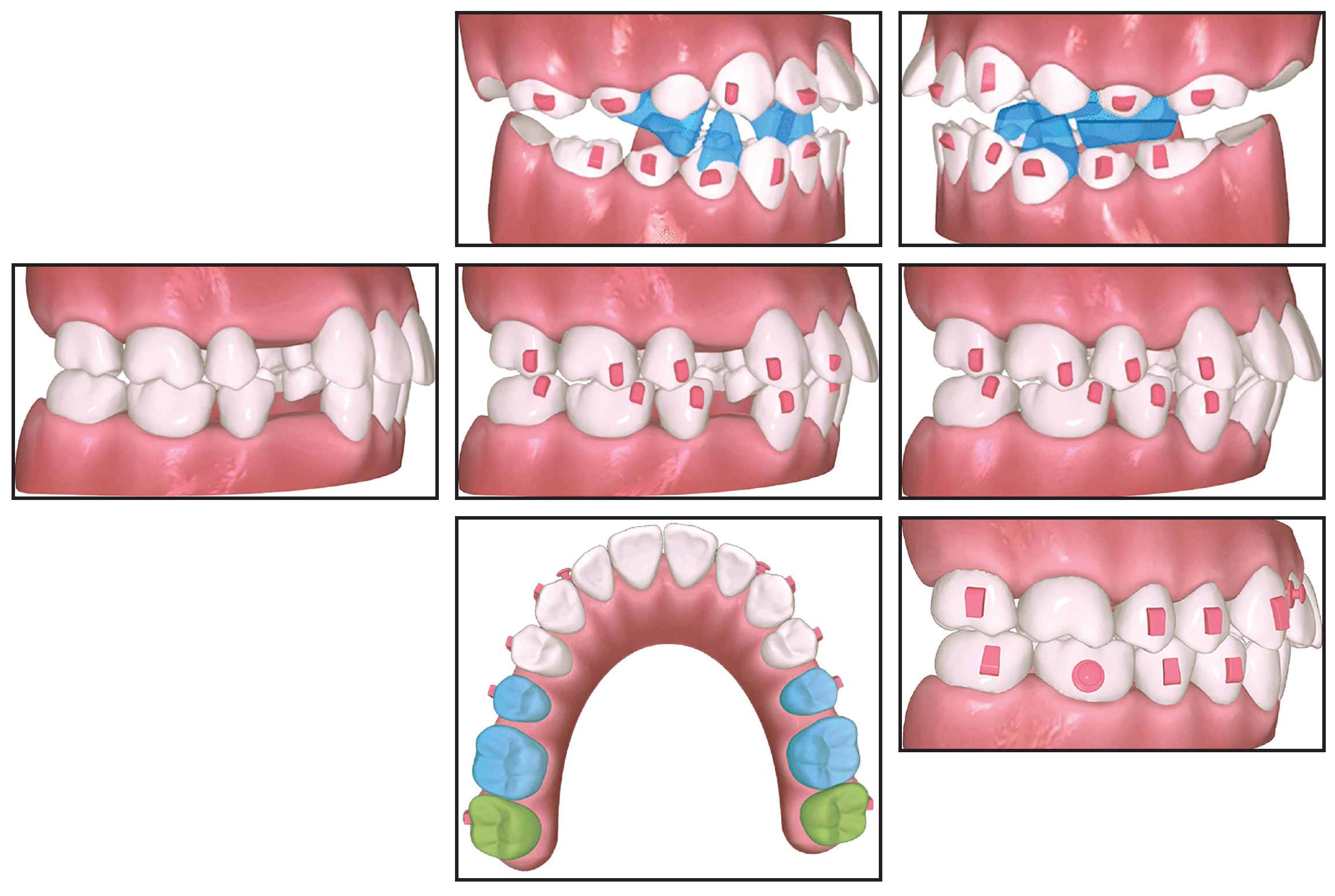
Fig. 1 Angel Aligners* A6 (top), A7 (middle), and A8 (bottom) protocols for mandibular advancement, premolar extraction, and molar distalization.
The A6 protocol, introduced in 2015, was the first among major clear aligner systems to incorporate occlusal blocks within the aligners, with the aim of replicating the functional and orthopedic effects of traditional twin block appliances. Specifically developed for growing patients, the integrated functional module is based on more than a decade of clinical use.
The A7 protocol applies controlled biomechanics for effective management of extraction space closure. Key components include anchorage preparation to mitigate the bowing effect, anterior torque control, and canine positioning to support ideal intercuspation and root angulation.
The A8 protocol employs a coordinated strategy involving customized staging, targeted attachments, and a distinct aligner trimline to optimize force vectors and minimize unwanted side effects. This approach enhances the predictability and efficiency of molar distalization, thus reducing the length of treatment.
Design Features
Two proprietary thermoplastic materials are employed to optimize Angel Aligner’s biomechanical performance. The first is MCS, a third-generation multilayer composite material introduced in 2020, which offers enhanced elastic recovery and sustained force delivery (Fig. 2). This formulation builds on prior materials, beginning with polycarbonate and PETG (first generation), followed by thermoplastic polyurethane (TPU) and coated PETG (second generation). In addition to MCS, the Pro product line includes masterControl (MC), a high-performance monolayer TPU material. The availability of two materials allows clinicians to tailor treatment based on specific mechanical requirements, such as stiffness, elasticity, and force decay, expanding the capabilities of aligner-based therapy.1
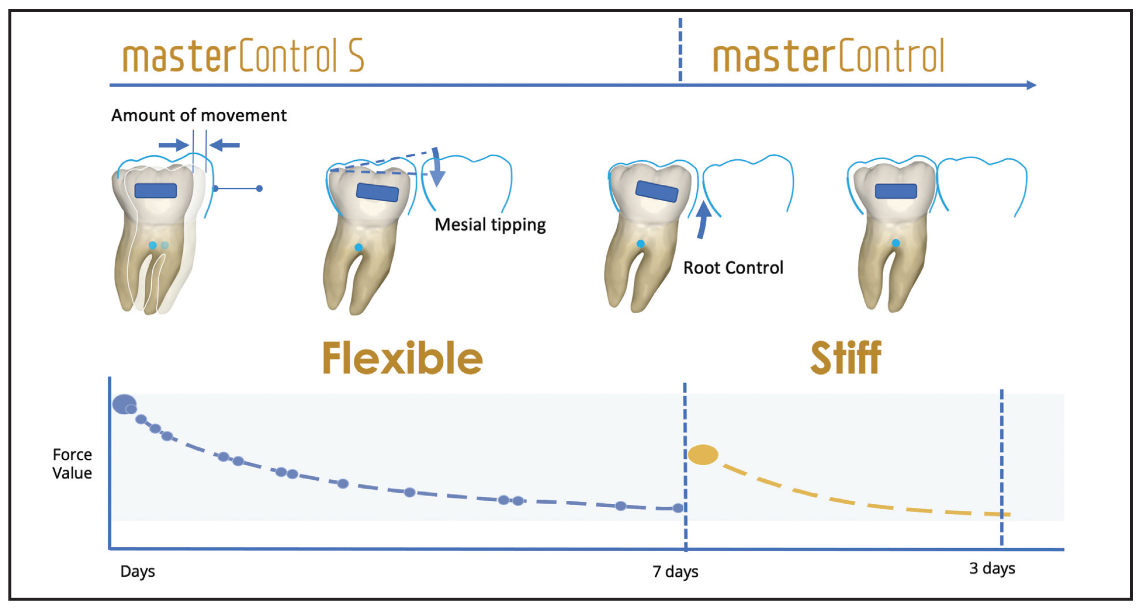
Fig. 2 masterControl S and masterControl materials used for staged force delivery during treatment.
Angel Aligner offers three distinct trimline configurations, two of which extend beyond the gingival margin to improve aligner engagement with the dentition and surrounding soft tissues, enabling more effective and predictable force transmission (Fig. 3).

Fig. 3 Trimline configurations, including extended gingival coverage for increased retention.
A customized half-wrap upper second molar design reduces distal resistance and thus improves force transfer during staged distalization, while minimizing undesirable side effects (Fig. 4).1
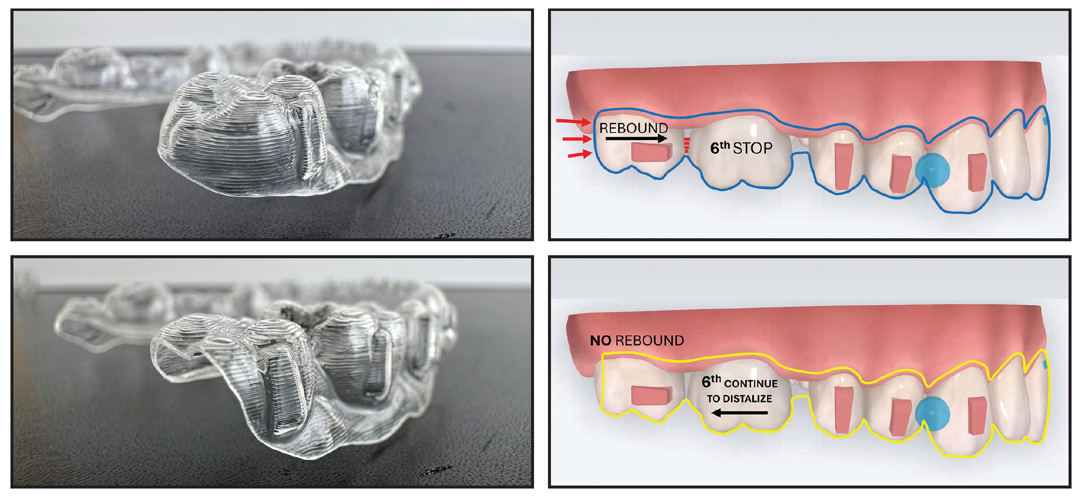
Fig. 4 Half-wrap maxillary second molar design used to reduce distal resistance and improve force transfer during staged distalization.
The Angel Aligner system also incorporates functional design elements aimed at addressing orofacial habits and optimizing muscle function during treatment. Among these features are tongue positioners and tongue spurs, which are integrated directly into the aligners (Fig. 5). The tongue positioner promotes proper tongue posture, while the tongue spurs are designed to discourage oral habits such as thumbsucking and tongue thrusting. Both components contribute to the neuromuscular reeducation needed for successful orthodontic outcomes and long-term treatment stability.1
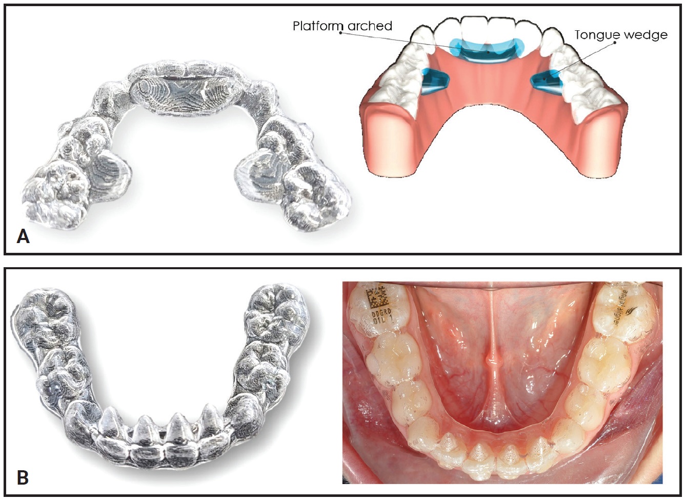
Fig. 5 Functional features incorporated into aligners. A. Passive tongue positioner. B. Tongue spurs that can be added to upper or lower incisors.
Among its mechanical features, Angel Aligner includes the AngelButton,* a prefabricated component that is integrated into the aligner surface, eliminating the need for chairside bonding of traditional buttons (Fig. 6).4 This design preserves the aligner’s active surface area, which is critical for effective force application. In contrast, conventional methods such as cutouts or bonded buttons can hinder mechanical efficiency and increase the risk of soft-tissue irritation. The AngelButton offers 360° omnidirectional flexibility, allowing elastic traction to be applied from labial, buccal, lingual, or interproximal positions for effective Class II or III correction, midline alignment, or occlusal adjustment. This provides a wide range of traction vectors without distorting the aligner trays, extending the initial biomechanical concept of aligner therapy from “pushing” to “pulling.”

Fig. 6 Prefabricated elastic attachment (AngelButton*) allowing multidirectional elastic traction.
Digital Integration
The Angel Aligner system is compatible with all intraoral scanners. Its iOrtho* treatment planning platform allows direct integration with many widely used systems, including 3Shape,*** Medit,† Alliedstar,†† and Dental Monitoring’s Smart STL,§ so that scan data can be transmitted directly without the need for manual file transfers.1
Among the digital tools in the iOrtho platform is iCeph, an AI-powered cephalometric analysis system designed to improve diagnostic speed and accuracy. The iCeph program provides fully automated cephalometric tracing and analysis in about five seconds, using the same software environment as for aligner treatment planning (Fig. 7).
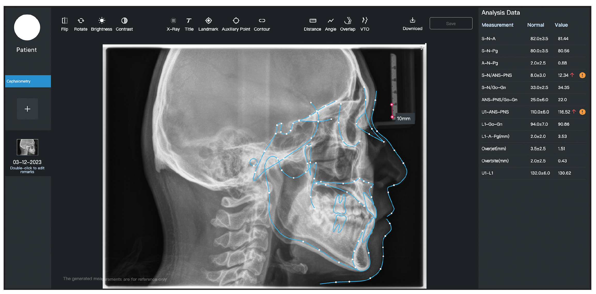
Fig. 7 Automated cephalometric tracing using iCeph* module.
In addition to initial diagnostics, iCeph supports superimposition of cephalometric tracings and can be used to estimate the skeletal maturity of growing patients (Fig. 8).
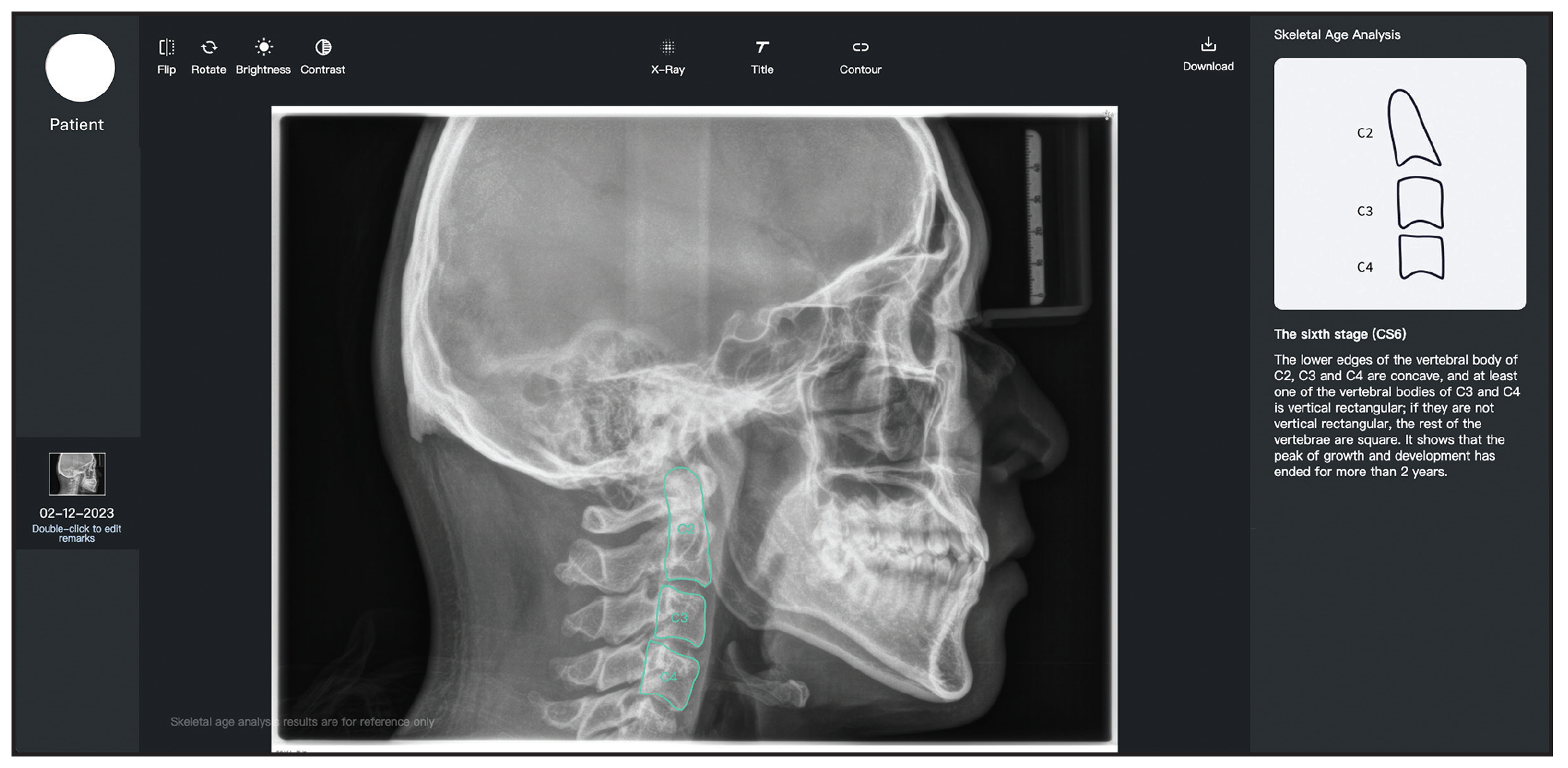
Fig. 8 Cephalometric superimposition and skeletal maturity assessment within iOrtho* platform.
The iOrtho platform also supports integration with CBCT data, enabling precise 3D control and visualization of bone, crown, and root positions within the planning process (Fig. 9).
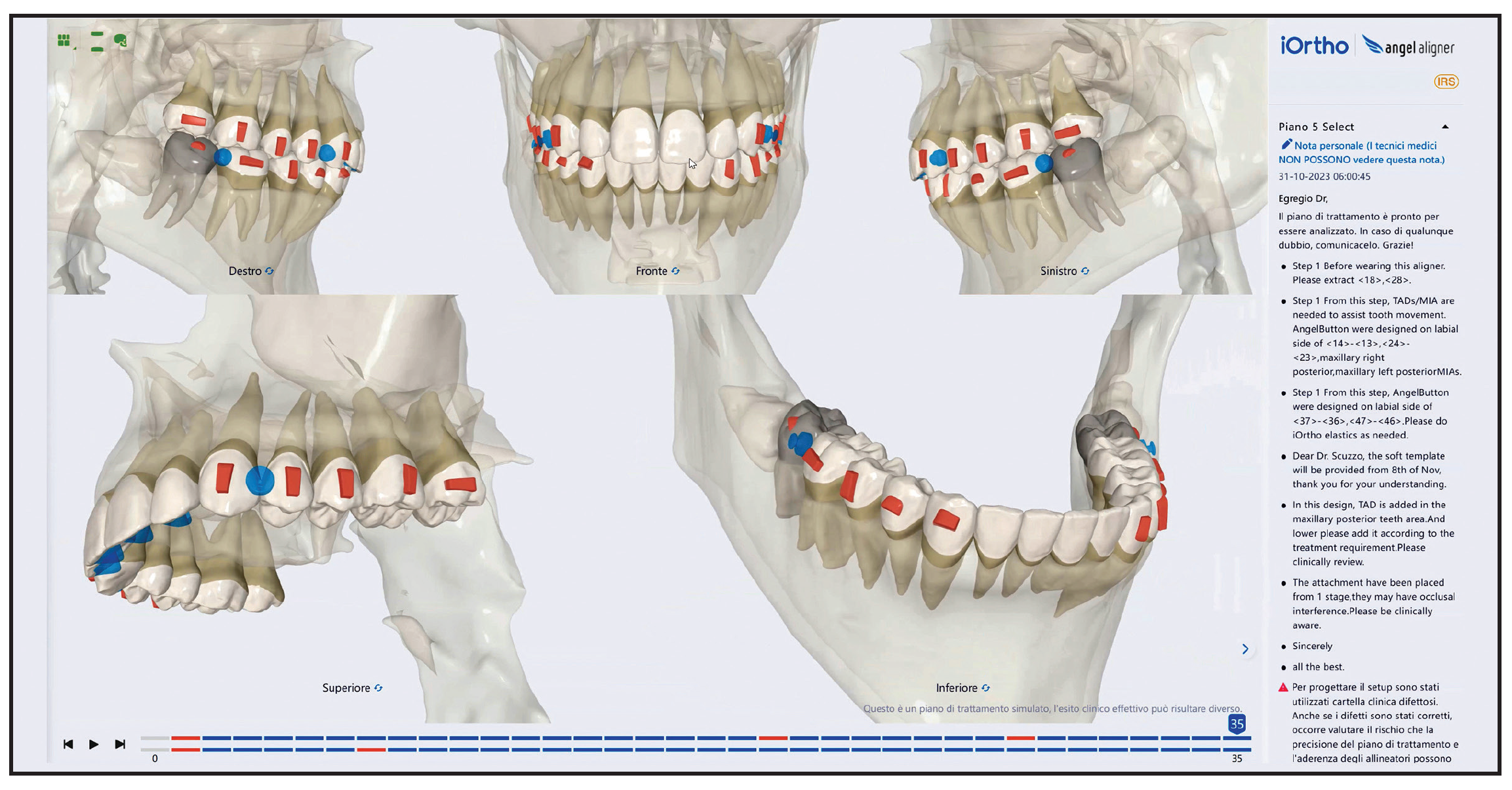
Fig. 9 Integration of CBCT data for visualization of crowns, roots, and surrounding bone structures.
The virtual Roots 2.0 is an advanced feature of the iOrtho platform designed to enhance root visualization in orthodontic treatment planning. Using a patented algorithm, it generates virtual root representations from standard panoramic radiographs, offering a patient-specific alternative when CBCT is not feasible (Fig. 10). While it provides more detail than conventional library-based models, it is not a substitute for true CBCT analysis. Together, the three available visualization modes—library-based roots, radiograph-derived virtual roots, and CBCT-based roots—can equip the clinician with a comprehensive set of diagnostic options for greater flexibility and integration into diverse digital workflows.1
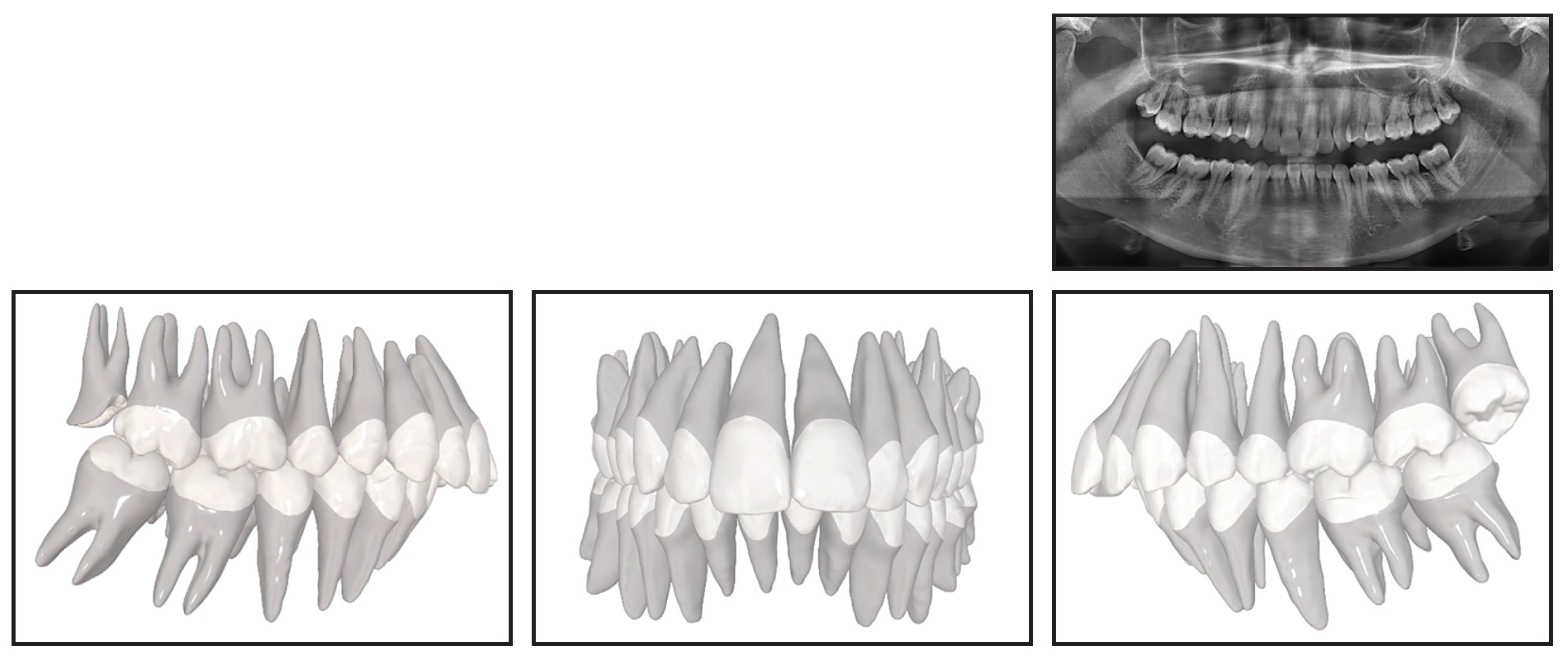
Fig. 10 Virtual Roots* 2.0 root estimation generated from panoramic radiograph data.
Case Report
A 24-year-old female presented with the chief complaint of upper anterior crowding (Fig. 11).
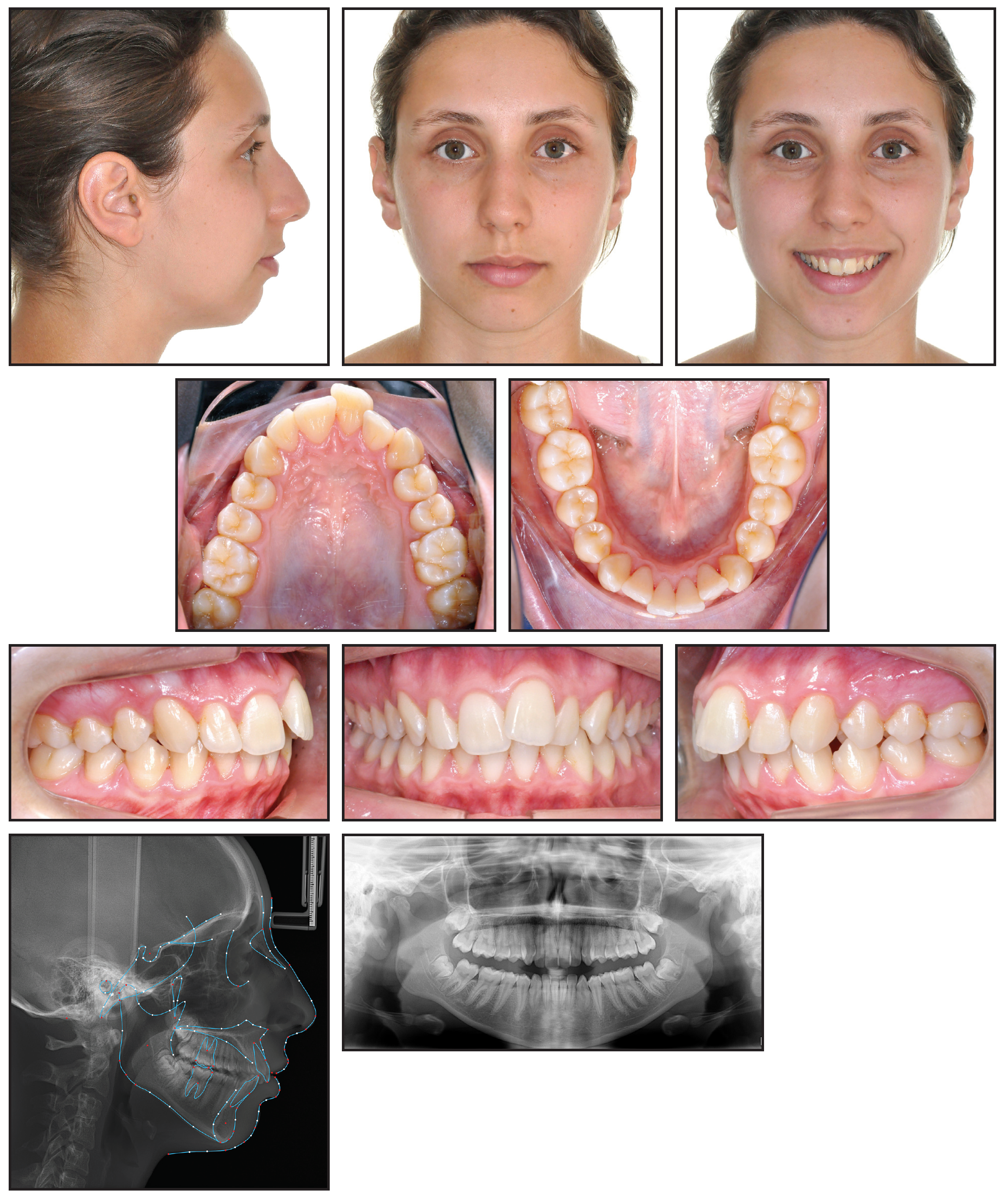
Fig. 11 24-year-old female patient with Class II, division 1 malocclusion before treatment.
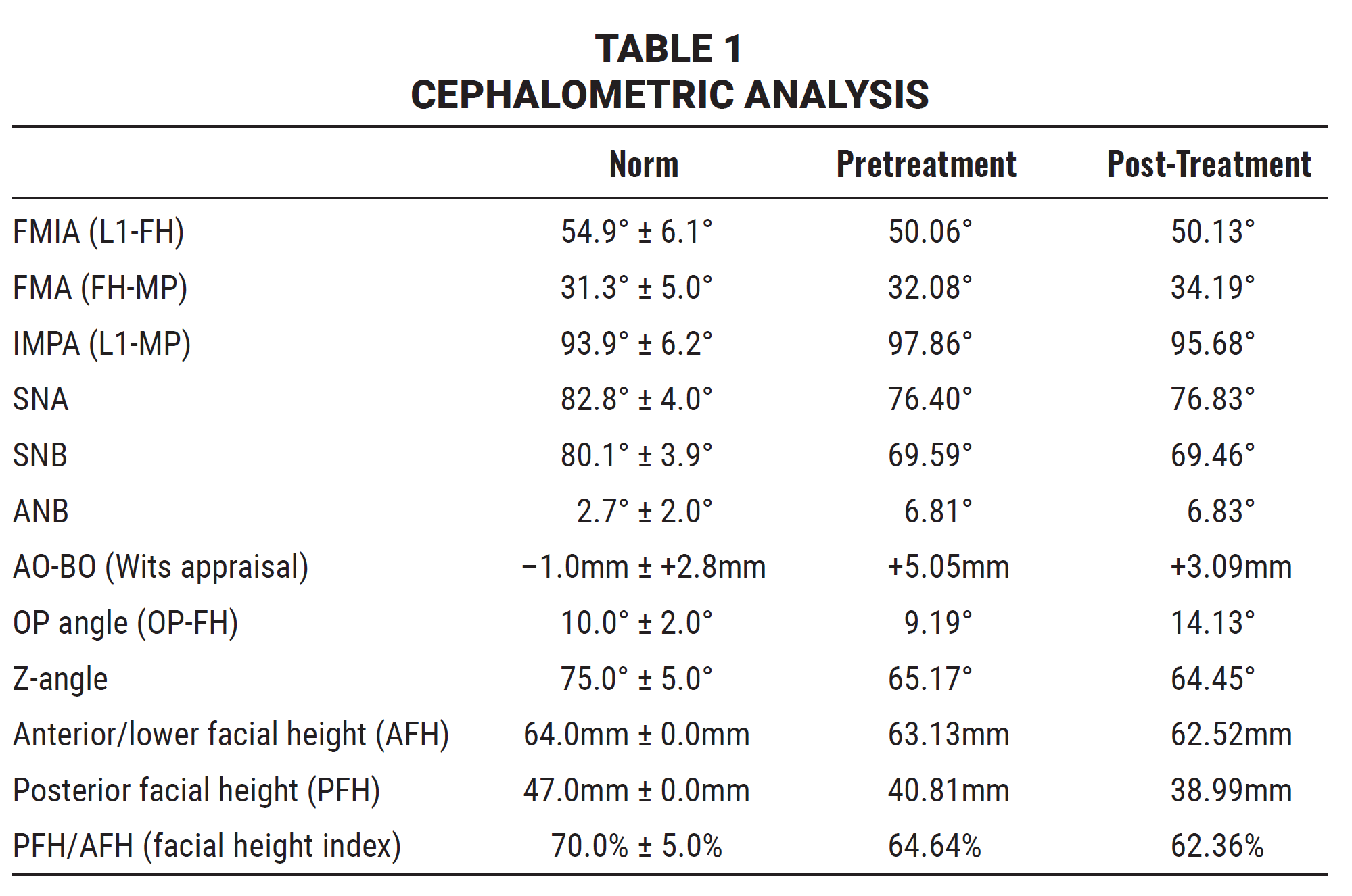
She had a weak Class I relationship on the right side and a Class II relationship on the left, with excessive overjet. Moderate crowding was present in both arches, and the entire upper left quadrant had migrated mesially. Panoramic radiography confirmed a full dentition with no bone defects, periapical lesions, or signs of TMD; all four third molars were present. Cephalometric analysis (Table 1) indicated a skeletal Class II relationship (ANB = 6.81°) and a normodivergent facial pattern tending toward hyperdivergence (IMPA = 97.86°). The diagnosis was a dental Class II, division 1 malocclusion with the upper segments positioned anteriorly.
Because of the mandibular retrusion, the ideal plan would have involved surgical-orthodontic treatment. When the patient refused surgery, a compromise solution was devised using Angel Aligner’s iOrtho digital platform.
Treatment objectives were to obtain Class I molar and canine relationships on both sides, with ideal overjet and overbite and centered facial and dental midlines. The treatment plan called for unilateral molar distalization on the left side using the A8 protocol, in conjunction with Class II elastics for distal traction and anterior bite ramps to disarticulate the arches. No overcorrection was applied.
The iOrtho system generated a treatment plan consisting of 31 aligner stages (Fig. 12). Aligners were to be changed every two weeks, as recommended for bodily molar distalization.5 The third molars were left in place to avoid bone volume issues during the application of distalization mechanics.
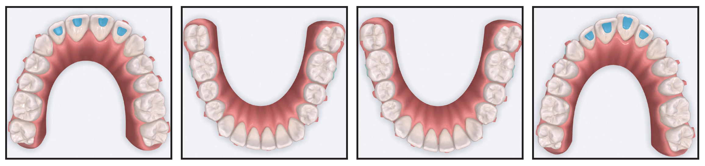
Fig. 12 Digital treatment setup, showing 31-stage distalization sequence.
In this case, the Class II elastics were attached to buttons bonded directly to the tooth enamel. This method not only increased anchorage for the distalization mechanics, but also exploited the expected side effects of the elastics to promote distal tipping of the upper canines and derotation of the lower molars (Fig. 13).6,7
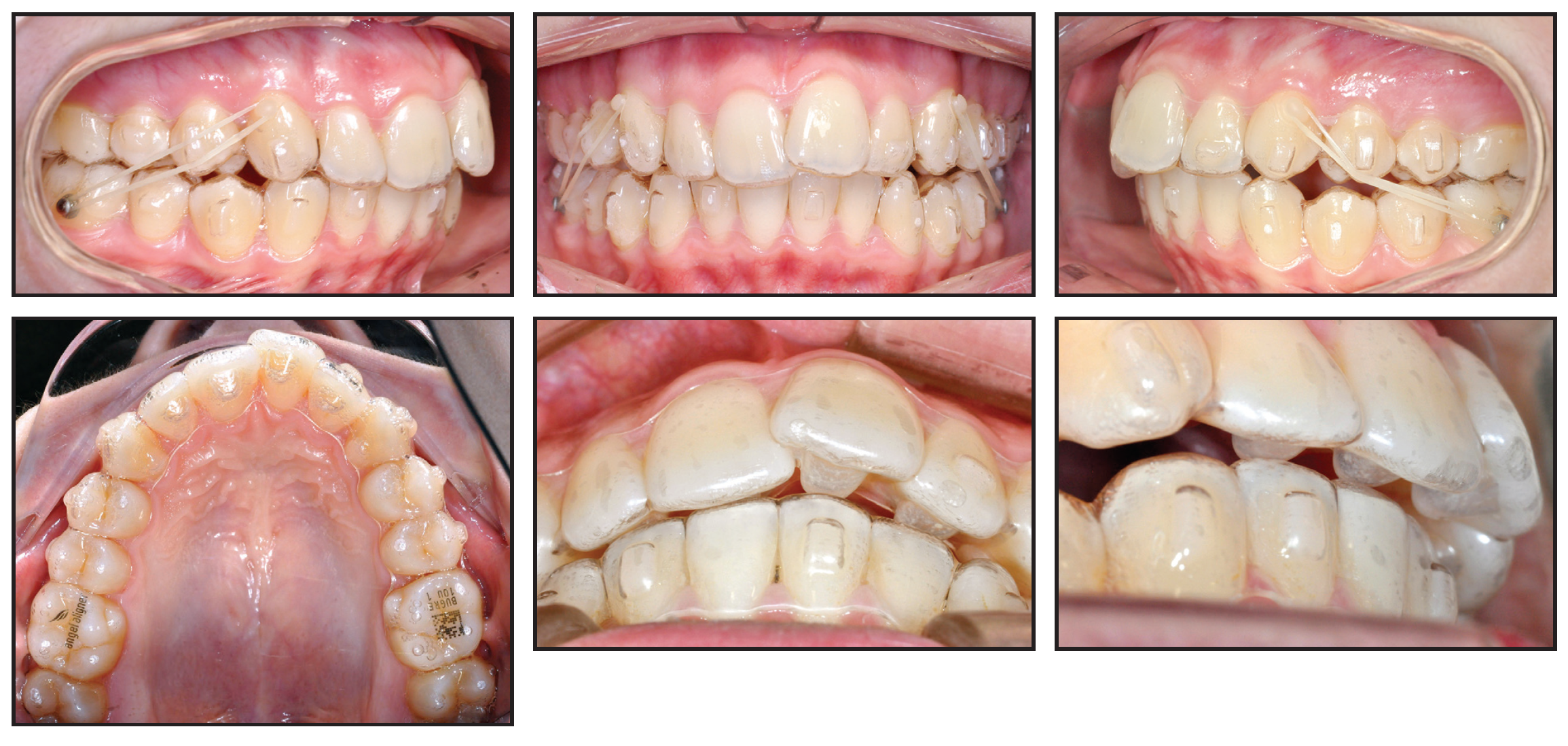
Fig. 13 After eight months of treatment, showing unilateral distalization mechanics with Class II elastics.
In 15 months of orthodontic treatment, the occlusal objectives were achieved in accordance with the initial digital plan (Fig. 14A and 14B).
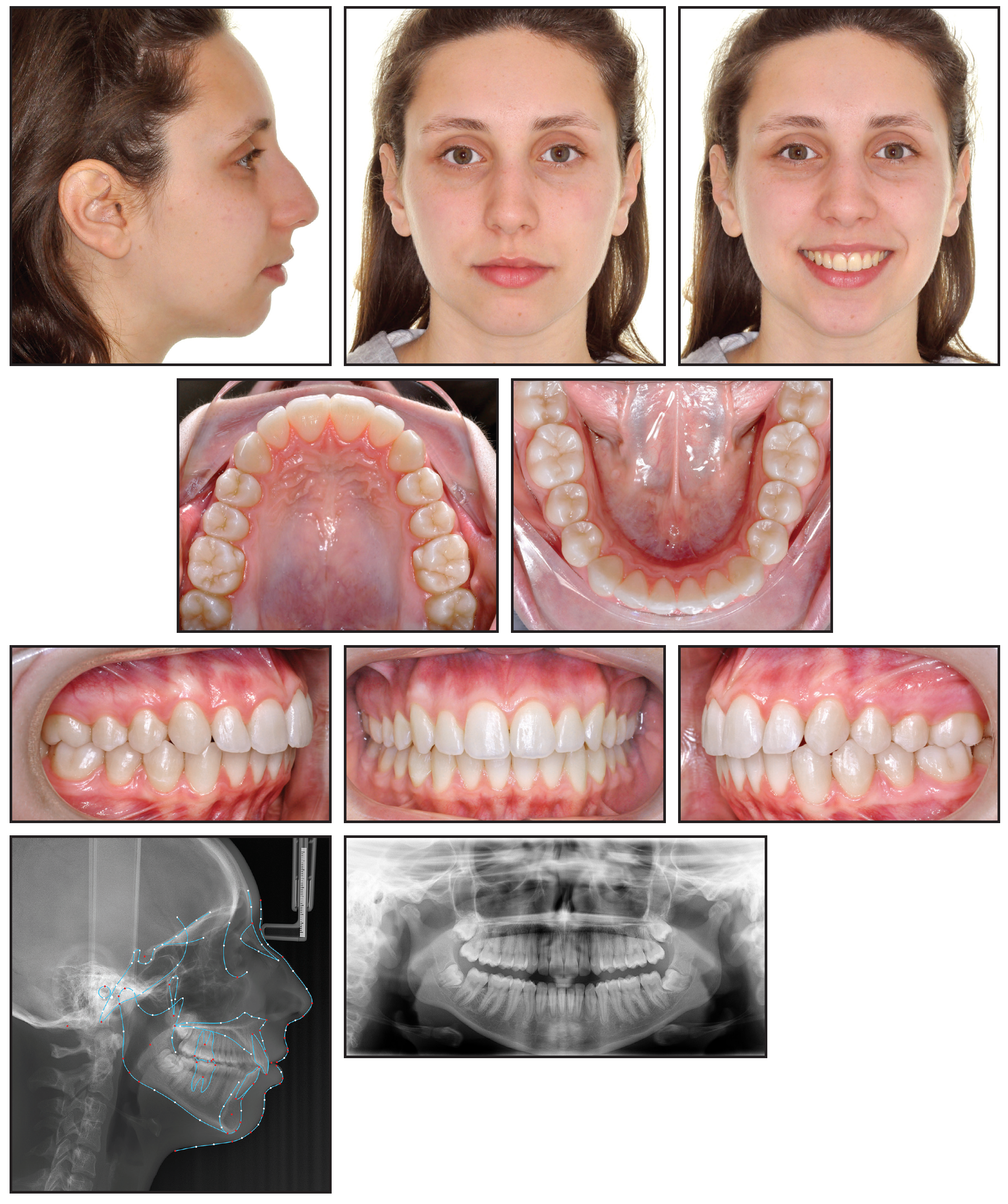
Fig. 14 A. Patient after 15 months of treatment (continued in next image).
The results included Class I molar and canine relationships on both sides, resolution of the upper and lower crowding, centering of the dental midlines on the facial midline, ideal overjet and overbite, and a fair degree of upper and lower dental expansion (Table 1). There was a consequent reduction in the buccal black corridors, and the patient finished with a wider smile.
Discussion
Unilateral molar distalization presents biomechanical challenges for clear aligner therapy, because of the need for anchorage preservation and control of such side effects as molar tipping, extrusion, and anterior proclination.8,9 In the present case, the Angel Aligner A8 protocol, supported by Class II elastics and optimized attachment design, facilitated controlled and predictable molar movement while minimizing unwanted effects.
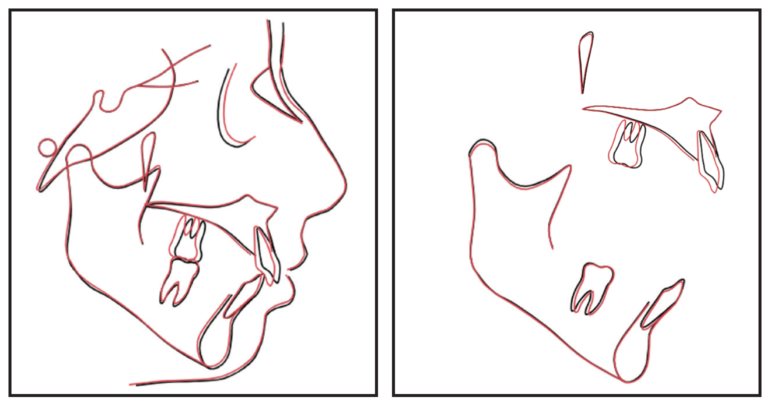
Fig. 14 (cont.) B. Superimposition of patient's pretreatment (black) and post-treatment (red) cephalometric tracings.
In the Angel Aligner system, tooth movement occurs progressively (typically in eight stages), with sequential distalization of premolars, canines, and molars using MC materials for controlled movement and AngelButtons* for flexible elastic traction. This system can produce 2-3mm of molar distalization with minimal tipping (1-2°)—in contrast to the increased inclination seen in other approaches. The half-wrap design for upper second molars can achieve superior distalization (95.5-96.5%) with minimal molar mesialization (.17mm), as compared to conventional full-wrap (.41mm) and half-wrap (.60mm) second molar designs.10 Reduced coverage of the most distal molars decreases resistance, allowing more effective force transmission to the premolars. This facilitates controlled and predictable distalization while reducing the risk of relapse for both first and second molars.
Launched as a cloud-based case management system, iOrtho has evolved from a platform for realistic 3D models and user-friendly interfaces to an AI-integrated tool for orthodontic mechanical simulations (masterForce) and immediate outcome predictions (the “Make It” feature), based on a vast orthodontic database.1 Since 2020, AI integration has enhanced iOrtho’s diagnostic capabilities, thus reducing human errors, with an emphasis on more efficient digital workflows for clear aligner treatments. In the cephalometric field, the Intelligent Cephalometry System (ICS)* is an AI-powered module that automates the tracing of anatomical landmarks with machine learning algorithms, achieving detection rates of more than 93% within 2mm of error (clinically acceptable) and more than 78% within 1mm.1 Developed in 2023-2024, the Intelligent Root System (IRS) incorporates AI-based segmentation to improve the fusion of CBCT and intraoral scan data, providing precise 3D visualizations to predict and monitor tooth movements—including the risks of root resorption or bone collisions. Overall, the continued development of the iOrtho platform reflects a broader shift toward the integration of more digital diagnostics within clear aligner therapy.1
As demonstrated by the case shown here, Angel Aligner’s advanced material properties, biomechanical protocols, and digital planning tools can produce predictable and efficient outcomes even in complex treatments such as unilateral molar distalization. The A8 protocol, combined with Class II elastics and optimized attachment systems, enables controlled tooth movement, anchorage preservation, and the achievement of functional and esthetic treatment goals without the need for surgical intervention. As clear aligner therapy continues to evolve, Angel Aligners provide an additional treatment option for managing a wide range of malocclusions.
FOOTNOTES
- *Angelalign Technology Inc., Paris, France; www.angelaligner.com.
- **Align Technology, Inc., San Jose, CA; www.aligntech.com.
- ***3Shape, Copenhagen, Denmark; www.3shape.com.
- †Medit, Seoul, South Korea; www.medit.com.
- ††Alliedstar, Shanghai, China; www.allied-star.com.
- §Dental Monitoring America Inc., Austin, TX; www.dental-monitoring.com.
REFERENCES
- 1. Angelalign Technology Inc.: A8 Protocol Guidelines, 2025.
- 2. Cusimano, C.; McLaughlin, R.P.; and Zernik, J.H.: Effects of four first bicuspid extractions on facial height in high-angle cases, J. Clin. Orthod. 27:594-598, 1993.
- 3. Suarez, O.; Alvarado-Lorenzo, A.; Calzadilla-Suárez, E.; Scuzzo, G.; León-Valencia, J.; Colino-Paniagua, C.; Granero-Marín, J.M.; and Colino Gallardo, P.: Comparison of the predictability of dentoalveolar expansion with different aligner systems in orthodontics: A longitudinal clinical study in adult patients, Appl. Sci. 15:9074, 2025.
- 4. García-Marín, C.; Otero-Pregigueiro, A.; and Iglesias-Linares, A.: Predictability of mesiodistal movement of upper and lower molars with clear aligners: A systematic review, Angle Orthod. 95:563-571, 2025.
- 5. Li, Y.; Wu, M.; Xing, X.; Chen, Y.; Zhang, C.; Hu, J.; and Shi, C.: Effects of maxillary molar distalization using clear aligners during the mixed dentition and early permanent dentition with 3 types of Class II traction: A three-dimensional finite element analysis, BMC Oral Health 25:717, 2025.
- 6. Rossini, G.; Parrini, S.; Castroflorio, T.; Deregibus, A.; and Debernardi, C.L.: Efficacy of clear aligners in controlling orthodontic tooth movement: A systematic review, Angle Orthod. 85:881-889, 2015.
- 7. Liu, X.; Cheng, Y.; Qin, W.; Fang, S.; Wang, W.; Ma, Y.; and Jin, Z.: Effects of upper-molar distalization using clear aligners in combination with Class II elastics: A three-dimensional finite element analysis, BMC Oral Health 22:546, 2022.
- 8. Ravera, S.; Castroflorio, T.; Garino, F.; Daher, S.; Cugliari, G.; and Deregibus, A.: Maxillary molar distalization with aligners in adult patients: A multicenter retrospective study, Prog. Orthod. 17:12, 2016.
- 9. Qiang, R.; Zhang, H.; Xu, Y.; Zhang, X.; Jin, F.; Jin, Z.; Qin, W.; and Gao, J.: Accuracy of maxillary molar distalization with clear aligners in three dimensions: A retrospective study based on CBCT superimposition, Clin. Oral Investig. 29:138, 2025.
- 10. Zhang, Y.; Wang, P.; Jiang, C.; Liu, Y.; Ma, X.; and Fan, C.: Biomechanical effects of clear aligners with different distal coverage designs combined with Class II elastic traction for maxillary first molar distalization: A finite element study, BMC Oral Health 25:1033, 2025.
-
 DR. GIACOMO SCUZZO
 -
 DR. GIUSEPPE SCUZZO
 -
 DR. ALVARADO LORENZO
Dr. Giacomo Scuzzo is a Specialist in Orthodontics and Clinical Director, Eline Clinic, Rome, Italy. Giuseppe Scuzzo is a Specialist in Orthodontics, Department of Orthodontics, Università Cattolica del Sacro Cuore, Largo Francesco Vito 1, 00168 Rome, Italy; e-mail: g.scuzzo@eline.it. Dr. Alvarado Lorenzo is a Specialist in Orthodontics, Department of Oral Surgery, Universidad de Salamanca, Villalba, Spain. Dr. Giacomo Scuzzo is a paid speaker for Angelalign Technology.