PEARLS
An Efficient and Effective Technique for Uprighting Mesially Erupting Lower Second Molars
Orthodontists can never have enough techniques in their armamentaria for uprighting mesioangular lower second molars. In this Pearl, as an alternative to a Halterman appliance, the authors use a beta titanium U-loop extended distally from the first-molar band to upright an impacted second molar. Note the crimpable stops on the U-loop to prevent the wire from sliding through the auxiliary tubes.
NEAL D. KRAVITZ, DMD, MS
Associate Editor for Pearls
Similar articles from the archive:
An Efficient and Effective Technique for Uprighting Mesially Erupting Lower Second Molars
Lower second molars erupt mesially in 2-3% of orthodontic patients.1 Cantilever springs and segmental mechanics with loops2 have been proposed for uprighting; more recently, one to three mini-implants have been used to provide skeletal anchorage.1 We have developed a much simpler method of uprighting a mesially inclined lower second molar using a U-loop wire spring.
Technique
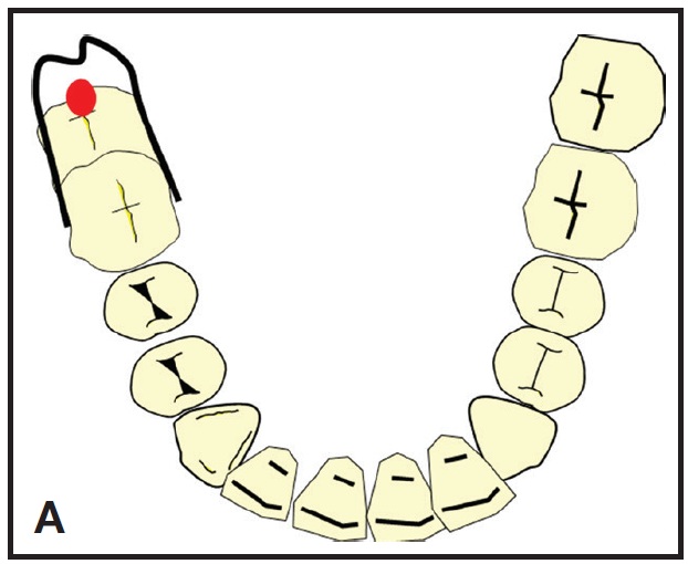
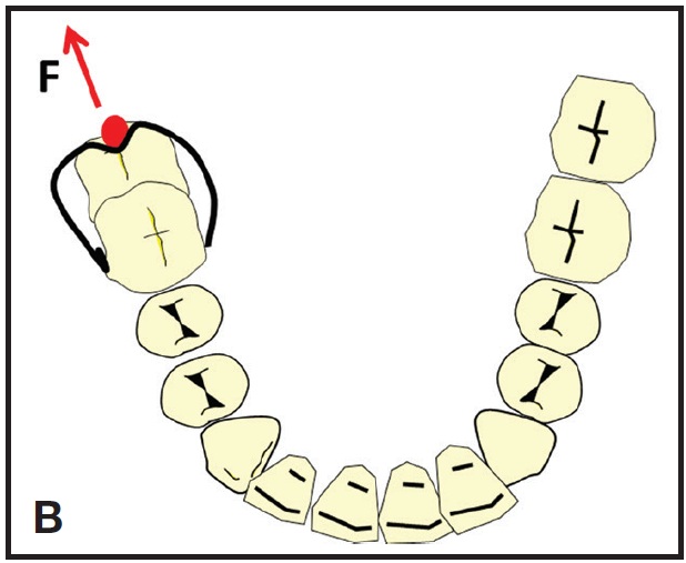
After surgical exposure of the impacted second molar with electrosurgery, a button is bonded to the occlusal surface. A U-loop is bent from .018" beta titanium wire and engaged between the buccal auxiliary tube and a lingually welded tube on the adjacent first molar. When placed passively in the tube, the loop should lie 2-3mm distal to the button (A). The center of the U is bent so that the wire will engage the button without displacement once the loop is activated (B). Crimpable stops are placed on the wire distal to the molar tube to prevent the loop from sliding after activation.
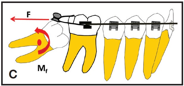
When the center of the U-loop is positioned under the button, a distal force is applied above the center of resistance of the second molar, generating a counterclockwise moment of the force (C). The U-loop can be further activated by repositioning the stops or rebonding the button more mesially.
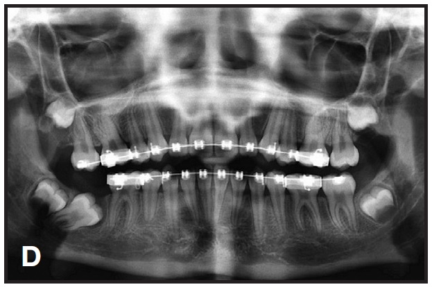
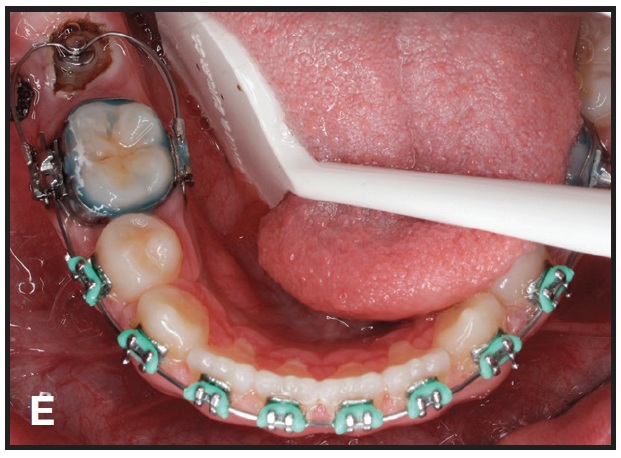
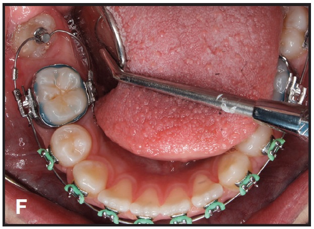
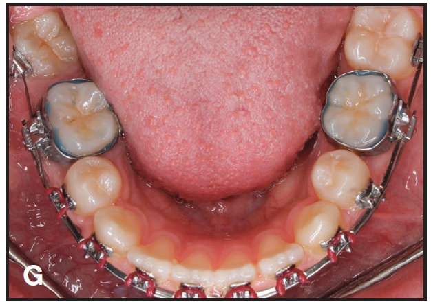
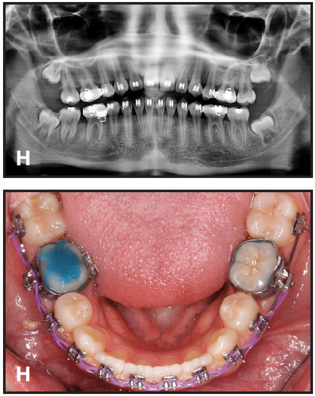
This technique is demonstrated in a 13-year-old female patient with an impacted lower right second molar (D). A U-loop spring was activated after surgical exposure of the tooth (E). Two months later, the second molar had been partially uprighted (F). After three more months of uprighting, the spring was removed and a continuous archwire was placed (G). It took another three months to completely upright the molar using straightwire mechanics, with no side effects on the anchor units (H).
REFERENCES
- 1. Mah, S.J.; Won, P.J.; Nam, J.H.; Kim, E.C.; and Kang, Y.G.: Uprighting mesially impacted mandibular molars with 2 miniscrews, Am. J. Orthod. 148:849-861, 2015.
- 2. Roberts, W.W. III; Chacker, F.M.; and Burstone, C.J.: A segmental approach to mandibular molar uprighting, Am. J. Orthod. 81:177-184, 1982.
-
 DR. JANAKIRAMAN
 -
 DR. HAKAMI
 -
 DR. URIBE
Assistant Professor Department of Orthodontics University of Louisville 501 S. Preston St. Louisville, KY 40202 nduggu@hotmail.com
Assistant Professor Division of Orthodontics Department of Preventive Dental Sciences College of Dentistry Jazan University Jazan, Saudi Arabia
Contributing Editor Journal of Clinical Orthodontics Associate Professor, Postgraduate Program Director, and Charles J. Burstone Endowed Professor Division of Orthodontics Department of Craniofacial Sciences University of Connecticut School of Dental Medicine Farmington, CT