


An Integrated AI Platform for Orthodontic Diagnosis and Treatment Planning
Uses for artificial intelligence (AI) have rapidly expanded within the orthodontic profession, offering new tools to improve diagnostic consistency, efficiency, and reproducibility.1,2 Applications such as automated cephalometric analysis, skeletal maturation assessment, cone-beam computed tomography (CBCT) segmentation, and digital model interpretation allow complex datasets to be analyzed in a more standardized and objective manner, without replacing the orthodontist’s clinical judgment.3-5
Beyond diagnosis, AI-driven systems are influencing treatment planning and decision-making. Machine-learning models can assist with extraction decisions, growth modification strategies, surgical case selection, and treatment simulation.6,7 When combined with three-dimensional imaging and digital workflows, these systems enable visualization of proposed tooth movements, estimations of treatment duration, and more effective communication of anticipated outcomes to patients.
This article introduces DR ANNIE (Data Research Artificial Neural Network Intelligence Expert), an end-to-end AI-assisted platform that integrates orthodontic diagnosis, treatment planning, and outcome prediction into a unified clinical workflow. The conceptual framework and diagnostic components of the system are described, followed by a clinical case illustrating its application in contemporary orthodontic practice.
Conceptual Framework of the DR ANNIE Platform
Rather than functioning as a single algorithm, the DR ANNIE platform operates as a modular architecture in which multiple AI engines process raw clinical data, generate structured diagnostic outputs, and support treatment sequencing (Fig. 1).
The workflow begins with the acquisition of standardized digital orthodontic records, including clinical photographs, intraoral scans, and panoramic and lateral cephalometric radiographs, as well as anteroposterior cephalometric and CBCT imaging when indicated. These inputs are processed through a series of specialized AI modules, each responsible for a specific diagnostic task such as landmark detection, skeletal classification, segmentation of dental and skeletal structures, and quantitative assessment of occlusal and anatomical relationships. Additional modules can be used to analyze clinical photographs for malocclusion, treatment needs, caries, periodontal disease, and restorations8; segment panoramic radiographs9; assess the TMJ10; and estimate case difficulty and treatment length.
Similar articles from the archive:
- THE CUTTING EDGE The Diagnostic Advantage of a CBCT-Derived Segmented STL Rendition of the Teeth and Jaws Using an AI Algorithm June 2021
- THE CUTTING EDGE Face Scanning and Digital Smile Design with an Intraoral Scanner March 2019
- MANAGEMENT & MARKETING The Future Is Now: How Millennials, Tech, and a Chess-Playing Computer Are Changing Orthodontics October 2017
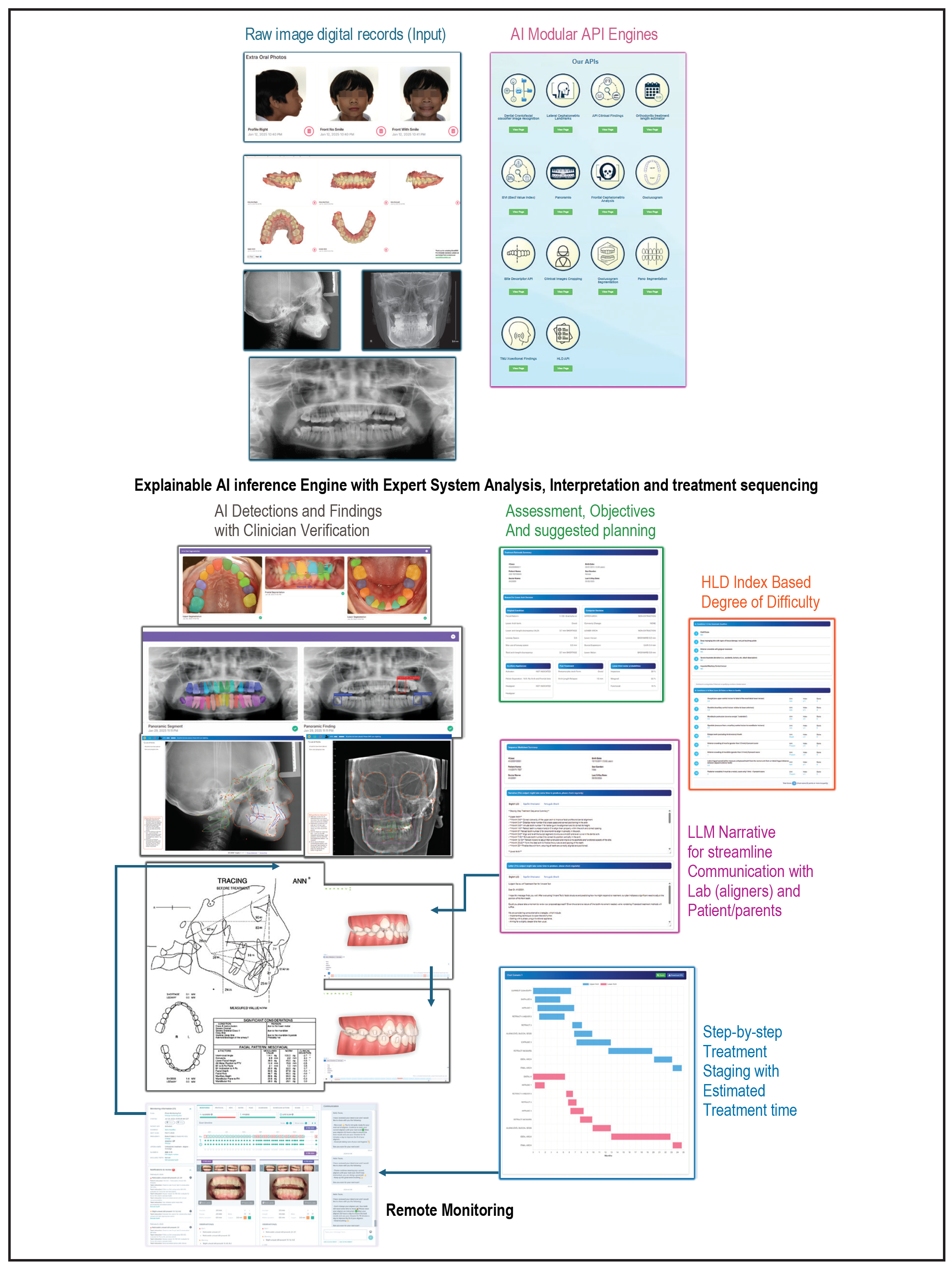
Fig. 1 Conceptual framework of DR ANNIE* platform. Raw digital records are processed through modular artificial intelligence (AI) engines; AI detections are verified by clinician before interpretation by explainable inference engine. Structured outputs include treatment objectives, staged planning with estimated treatment durations, and narrative summaries to support communication with laboratories and patients. API = application programming interface; HLD = Handicapping Labiolingual Deviation; LLM = large language model.
A defining feature of the platform is the integration of verification by the clinician at each critical stage of analysis. AI-generated detections and measurements are presented in a diagnostic review interface for the orthodontist to review, confirm, or modify before downstream interpretation occurs. This hybrid workflow ensures that diagnostic outputs will remain clinically valid while benefiting from the efficiency and consistency of automated analysis.
Following verification, diagnostic findings are processed by an explainable AI inference engine that combines machine-learning outputs with expert-system logic to assess treatment complexity, identify biomechanical constraints, and sequence treatment objectives. Difficulty levels can be graded using a modified Handicapping Labiolingual Deviation (HLD) index.11
The individual engines were trained on about 50,000 successfully treated orthodontic cases selected from a larger pool of about 200,000 completed cases, collected over nearly three decades from multiple clinicians using different treatment philosophies, biomechanical approaches, and appliances. These datasets included annotated clinical photographs, intraoral scans, cephalometric and panoramic radiographs, and longitudinal treatment records.
Several components of the platform have undergone independent or institutional validation, including lateral12 and CBCT-derived anteroposterior13 cephalometric landmark identification, cervical vertebral maturation assessment, treatment-length prediction,14 and automated case difficulty assessment. Landmark detection accuracy is reportedly in the range of 90-95%.12-16
AI Assessment, Objectives, and Suggested Treatment Planning
Following AI-assisted diagnosis, the DR ANNIE platform integrates imaging-based findings with patient age, sex, and race to evaluate extraction vs. nonextraction strategies, orthopedic vs. camouflage approaches, anchorage requirements, and surgical vs. nonsurgical options. Rather than generating a single prescriptive plan, the system presents treatment options, predicted outcomes, and biomechanical considerations to assist the orthodontist in selecting the most appropriate approach for each patient.
AI-assisted simulations allow the orthodontist to evaluate proposed tooth movements and identify biomechanical limitations before initiating treatment. These simulations also facilitate a comparison of alternative treatment strategies. Suggested staging includes wire sequencing and anchorage strategies for fixed appliances and tooth-movement staging for clear aligners.
By analyzing historical treatment datasets and patient-specific characteristics, the system also estimates treatment duration, identifies potential risk factors, and highlights areas requiring closer monitoring.14
A large language model (LLM)-based narrative generation layer translates diagnostic findings, treatment objectives, and staging information into narratives for orthodontic laboratories, referring providers, and patients or parents.
Integration with AI-driven remote monitoring systems enables longitudinal tracking of tooth movements and appliance performance throughout active treatment.17-19 The observed treatment progress can be compared with predicted outcomes, allowing the clinician to intervene early when deviations occur.
Case Report
A 12-year-old female presented with the chief complaint of crowded teeth. Clinical examination found a mesocephalic facial pattern and a straight profile, with no asymmetry. The patient exhibited moderate crowding in both arches, irregular incisor alignment, a 2mm overjet, a 1mm overbite, and a super-Class I molar relationship.
Pretreatment orthodontic records, including standardized extraoral and intraoral photographs (Fig. 2), intraoral digital scans, lateral and frontal cephalometric radiographs, and a panoramic radiograph, were uploaded to the DR ANNIE platform.
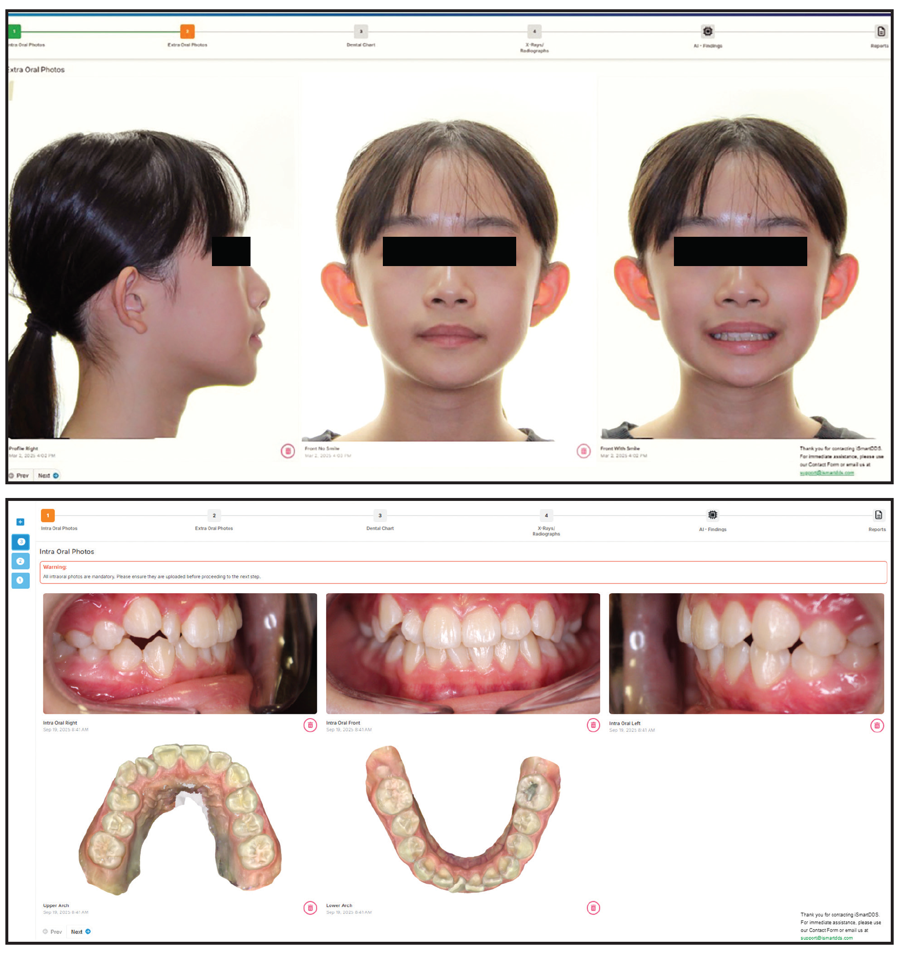
Fig. 2 Standardized extra- and intraoral pretreatment photographs of 12-year-old female patient with moderate crowding in both arches.
In the intraoral scans, individual teeth were automatically segmented and labeled, allowing a quantitative evaluation of archform, tooth-size distribution, and occlusal relationships (Fig. 3).
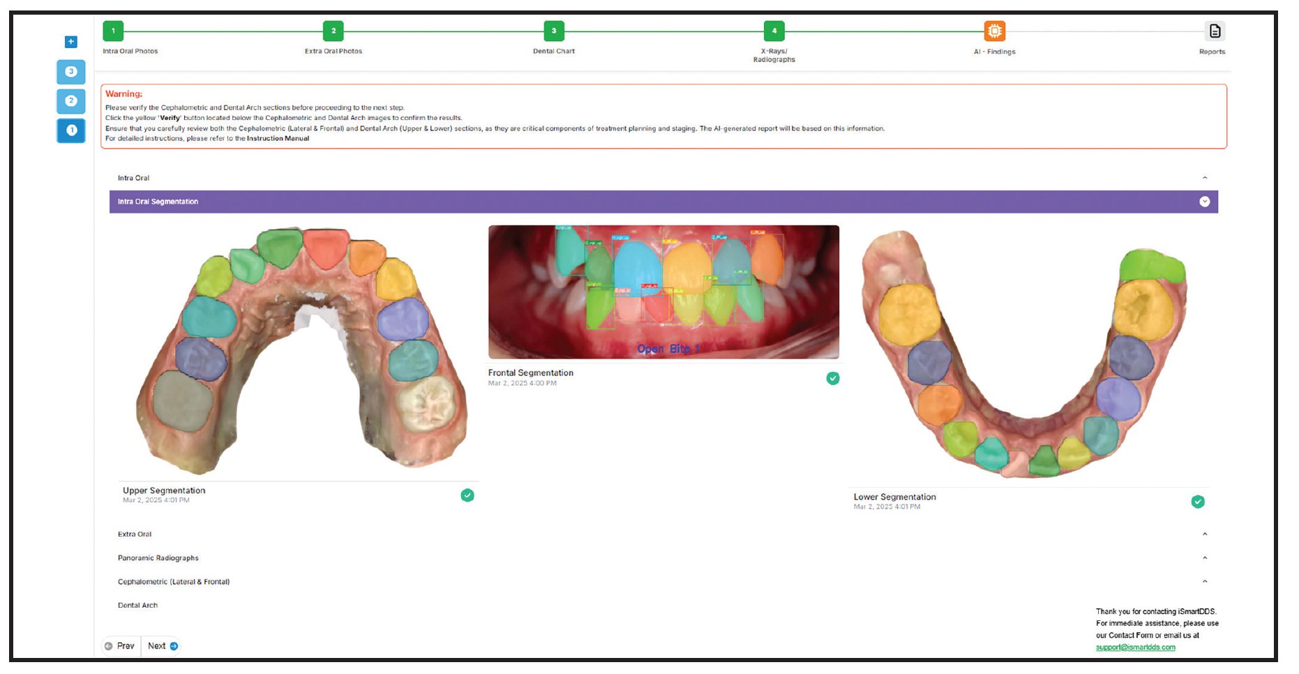
Fig. 3 AI-assisted intraoral scan segmentation of maxillary and mandibular arches with automated tooth identification and occlusogram analysis, as verified by clinician.
Lateral and frontal cephalometric radiographs were analyzed using AI-assisted landmark identification (Fig. 4), demonstrating a Class I skeletal pattern and a brachycephalic vertical pattern. Panoramic segmentation identified individual teeth and flagged potentially impacted third molars on both sides (Fig. 5).
The automated HLD index-based assessment yielded a total score of 14, indicating a low-to-moderate degree of treatment difficulty (Fig. 6). This supported the feasibility of a nonextraction treatment; the score did not meet the threshold typically required for public insurance coverage.
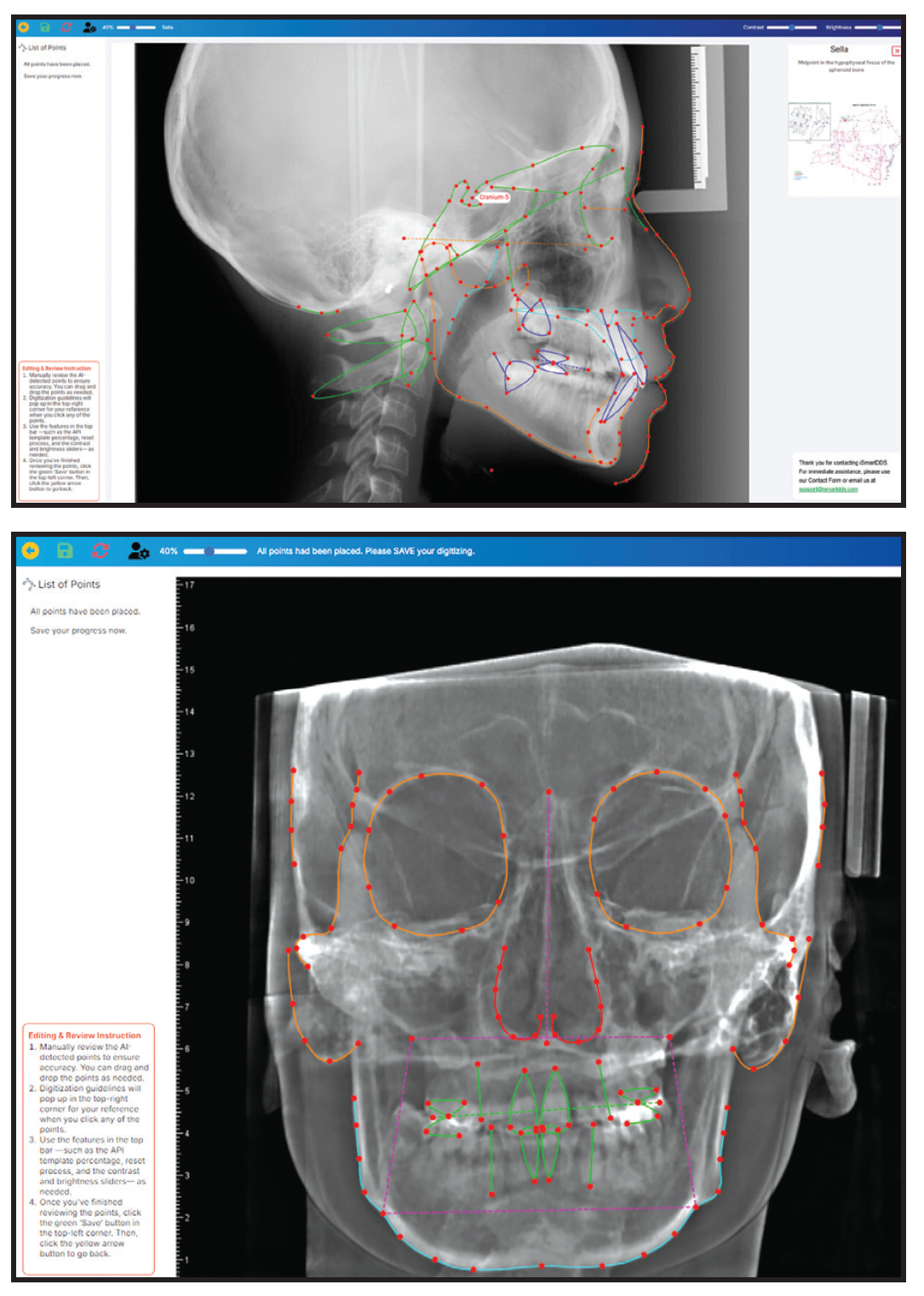
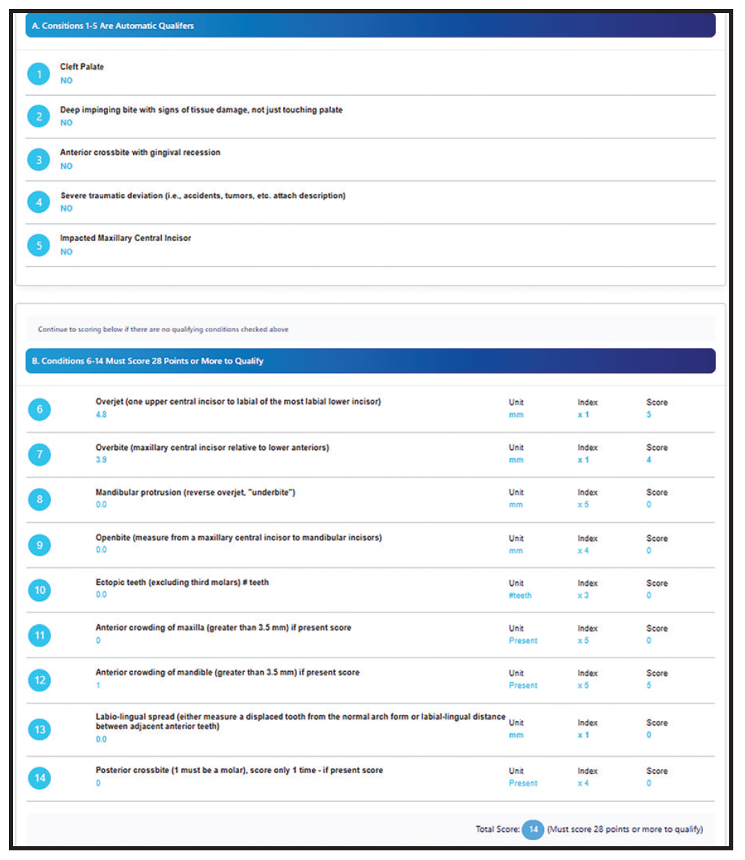
Fig. 6 Automated HLD index-based degree of difficulty assessment, generated from clinician-verified diagnostic inputs.
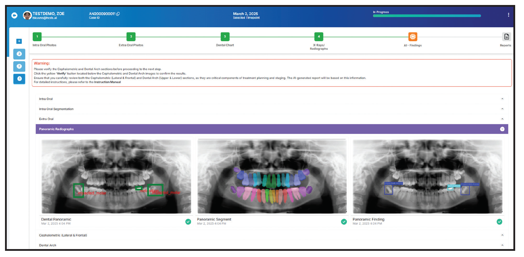
Fig. 5 AI-assisted panoramic segmentation and analysis, identifying individual teeth and impacted molars, as verified by clinician.
Key findings influencing treatment decisions included the growth pattern, an ovoid lower archform, a lower arch-length discrepancy of 3.7mm, an absence of leeway space, and a lack of indication for orthopedic or auxiliary appliances. The AI-supported decision analysis recommended a nonextraction approach in both arches, with space management achieved through minor posterior distalization, limited incisor repositioning, and transverse coordination with buccal segment alignment (Fig. 7A). Alternative approaches considered for this patient included limited extraction therapy and conventional fixed appliance therapy. The final decision was made by the treating orthodontist after considering the patient’s esthetics, growth pattern, and eruption status, as well as biomechanical feasibility. Clear aligner therapy was selected based on the doctor’s preference.
The platform then generated a step-by-step treatment staging sequence for each arch, with an estimated overall treatment duration of 16-17 months (Fig. 7B). In both arches, the early phases emphasized posterior tooth distalization and incisor vertical control, followed by buccal segment alignment and refinement of archform.
The platform’s LLM-based narrative translated technical staging into patient-friendly language, describing month-by-month objectives for each arch (Fig. 8). This narrative was reviewed and edited by the orthodontist before it was used in discussions with the patient and parents and for communication with the aligner laboratory.
The clinician-approved staging was exported to the aligner simulation (Fig. 9).
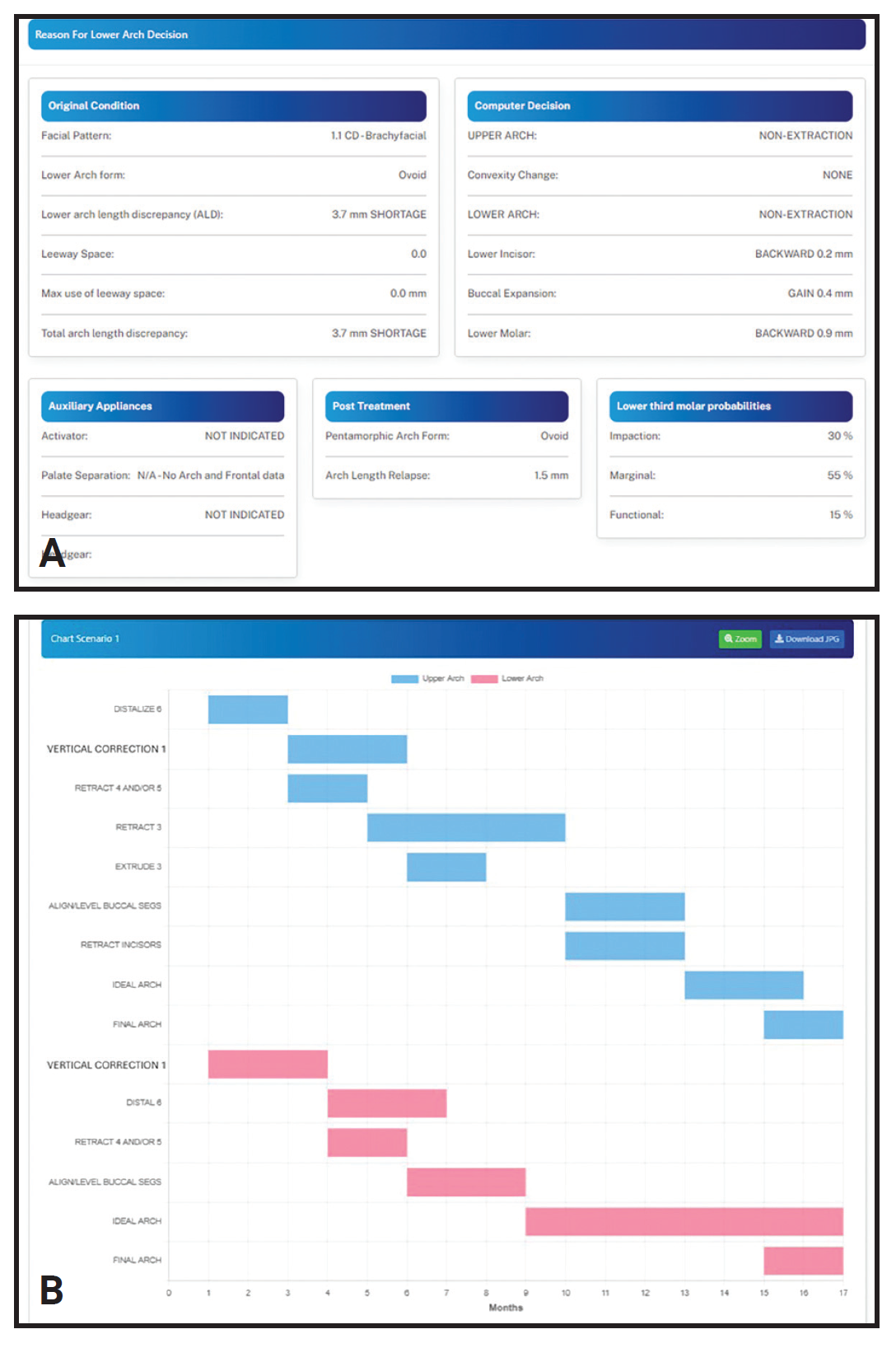
Fig. 7 A. Structured AI-generated treatment objectives and decision analysis, supporting nonextraction treatment approach in both arches. B. AI-guided step-by-step treatment staging for maxillary and mandibular arches, with estimated treatment durations.
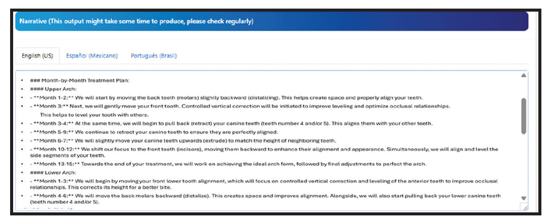
Fig. 8 LLM-generated narrative summary translating treatment staging into patient- and parent-friendly language, after review by clinician.
After 11 months of clear aligner therapy, the observed tooth positions corresponded closely with the predicted stages (Fig. 10).
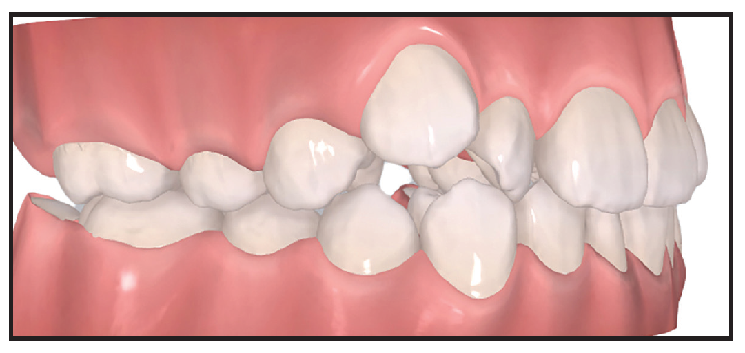
Fig. 9 Three-dimensional, AI-guided clear aligner treatment simulation, showing initial tooth positions and planned orthodontic movements based on clinician-approved staging.
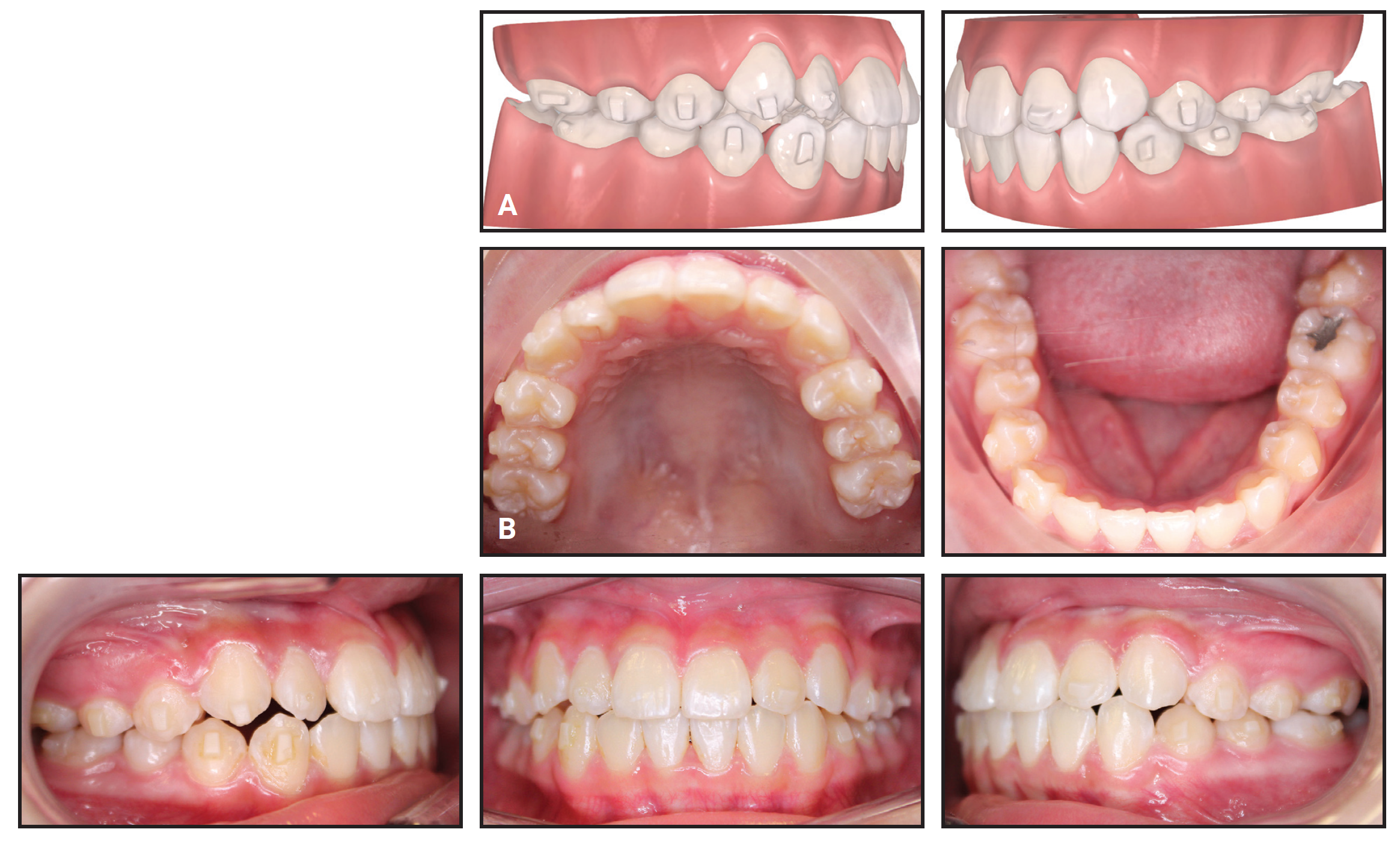
Fig. 10 A. Intermediate 3D simulation, showing progress of dental alignment and occlusal correction during clear aligner therapy. B. Patient after 11 months of clear aligner treatment.
Discussion
AI platforms such as DR ANNIE are designed to integrate into existing orthodontic workflows. In routine practice, they can be introduced at the diagnostic and consultation stages using records that meet the established standard of care. By automating time-intensive analytical tasks, AI enables orthodontists to focus more on clinical decision-making, patient communication, and treatment execution. AI systems may also reduce variability between providers within group practices or multilocation clinics.
Visual representations of malocclusion severity, treatment objectives, and step-by-step staging, reinforced by clinician-reviewed narratives, allow patients and parents to better understand the rationale behind recommended treatment. This improved understanding has the potential to increase case acceptance, help manage expectations, and improve compliance.
We recommend gradual adoption of an AI platform, beginning with diagnostic assistance and case visualization before progressing to treatment staging and outcome prediction. Such an approach will enable the orthodontist and staff to build trust in the system while preserving effective workflows.
AI-based systems do have certain limitations. First, their detections and recommendations can be inaccurate and must be verified and interpreted by the orthodontist. In addition, although several individual components of DR ANNIE have been validated, the fully integrated system has yet to undergo an end-to-end validation.
As AI models continue to evolve, further applications are expected to include more refined outcome prediction, integration of longitudinal treatment data, and personalized risk assessment. The most effective future applications of AI in orthodontics will be those that can augment clinical expertise, enhance patient-centered care, and support evidence-based decision-making, without replacing the clinician’s judgment.
FOOTNOTES
- *Trademark of ismartoffice.com, Chicago, IL; www.ismartoffice.com.
REFERENCES
- 1. Choi RY, Coyner AS, Kalpathy-Cramer J, Chiang MF, Campbell JP. Introduction to machine learning, neural networks, and deep-learning. Transl Vis Sci Technol. 2020;9(2):14. doi: https://www.doi.org/10.1167/tvst.9.2.14
- 2. Russell SJ, Norvig P. Artificial Intelligence: A Modern Approach, Global Edition. 3rd ed. Pearson; 2016.
- 3. Kunz F, Stellzig-Eisenhauer A, Zeman F, Boldt J. Artificial intelligence in orthodontics: evaluation of a fully automated cephalometric analysis using a customized convolutional neural network. J Orofac Orthop. 2020;81(1):52-68. doi: https://www.doi.org/10.1007/s00056-019-00203-8
- 4. Atici SF, Ansari R, Allareddy V, Suhaym O, Cetin AE, Elnagar MH. Fully automated determination of the cervical vertebrae maturation stages using deep learning with directional filters. PLoS One 2022;17(7):e0269198. doi: https://www.doi.org/10.1371/journal.pone.0269198
- 5. Cui Z, Fang Y, Mei L, et al. A fully automatic AI system for tooth and alveolar bone segmentation from cone-beam CT images. Nat Commun. 2022;13(1):2096. doi: https://www.doi.org/10.1038/s41467-022-29637-2.
- 6. Taraji S. Novel Machine Learning Algorithms for Prediction and Treatment Decision in Adult Patients with Class III. Thesis. University of Illinois Chicago; 2022. https://www.doi.org/doi: 10.25417/uic.20254257
- 7. Rhee M, Elnagar MH, Allareddy V, Milani OH, Cetin AE, Sanchez FJ. Integrating machine learning for treatment decisions in anterior open bite orthodontic cases: a retrospective study. J World Fed Orthod. 2025;14(3):140-147. doi: https://www.doi.org/10.1016/j.ejwf.2024.12.001
- 8. Talaat S, Kaboudan A, Talaat W, et al. The validity of an artificial intelligence application for assessment of orthodontic treatment need from clinical images. Semin Orthod. 2021;27(2):164-171. doi: https://www.doi.org/10.1053/j.sodo.2021.05.012
- 9. Wirtz A, Mirashi SG, Wesarg S. Automatic teeth segmentation in panoramic X-ray images using a coupled shape model in combination with a neural network. In: Frangi A, Schnabel J, Davatzikos C, Alberola-López C, Fichtinger G, eds. Medical Image Computing and Computer Assisted Intervention—MICCAI 2018. Lecture Notes in Computer Science. Vol. 11073. Springer Verlag; 2018:712-719. doi: https://www.doi.org/10.1007/978-3-030-00937-3_81
- 10. Talaat WM, Shetty S, Al Bayatti S, et al. An artificial intelligence model for the radiographic diagnosis of osteoarthritis of the temporomandibular joint. Sci Rep. 2023;13(1):15972. doi: https://www.doi.org/10.1038/s41598-023-43277-6.
- 11. Makki A, Elnagar MH, Sanchez F, et al. Assessment of Handicapping Labio-Lingual Deviation index scoring methods and their effect on orthodontic treatment coverage by Medicaid. J Public Health Dent. 2022;82(4):478-483. doi: https://www.doi.org/10.1111/jphd.12505
- 12. Chwa E. AI-Based Cephalometric Landmark Detection via Deep Learning Networks Evaluation. Thesis. University of Illinois Chicago; 2025. https://indigo.uic.edu/articles/thesis/AI-Based_Cephalometric_Landmark_Detection_via_Deep_Learning_Networks_Evaluation/29338292
- 13. Kocher M. Assessment of Frontal Cephalogram Tracing Accuracy Generated by CNNs and Machine Learning. Thesis. University of Illinois Chicago; 2026. doi: https://www.doi.org/10.25417/uic.32993771.v1
- 14. Elnagar MH, Pan AY, Handono A, et al. Utilization of machine learning methods for predicting orthodontic treatment length. Oral. 2022;2(4):263-273. doi: https://www.doi.org/10.3390/oral2040025
- 15. Hatoum H, Talaat W, Kaboudan A, Hamed A, Mahmoud E, Talaat S, Shetty S, Sadek M. Maxillary crowding and spacing: validation of an artificial intelligence model vs. digitally assisted human observer. J Orofac Orthop. 2026 Jan 3. English. doi: https://www.doi.org/10.1007/s00056-025-00634-6
- 16. Elnagar MH, Pan AY, Handono A, et al. Artificial intelligence-powered prediction of orthodontic treatment duration: identifying short and prolonged cases from a dataset of more than 10,000 patients. J Orthod Sci. 2026;15(1). doi: https://www.doi.org/10.4103/jos.jos_186_25
- 17. Morris RS, Hoye LN, Elnagar MH, et al. Accuracy of dental monitoring 3D digital dental models using photograph and video mode. Am J Orthod Dentofacial Orthop. 2019;156(3):420-428. doi: https://www.doi.org/10.1016/j.ajodo.2019.02.014
- 18. Homsi K, Snider V, Kusnoto B, et al. In-vivo evaluation of artificial intelligence driven remote monitoring technology for tracking tooth movement and reconstruction of 3-dimensional digital models during orthodontic treatment. Am J Orthod Dentofacial Orthop. 2023;164(5):690-699. doi: https://www.doi.org/10.1016/j.ajodo.2023.04.019
- 19. Snider V, Homsi K, Kusnoto B, et al. Effectiveness of AI-driven remote monitoring technology in improving oral hygiene during orthodontic treatment. Orthod Craniofac Res. 2023; 26(suppl 1):102-110. doi: https://www.doi.org/10.1111/ocr.12666
-
 DR. ELNAGAR
-
 DR. SANCHEZ
-
 DR. SAMBATARO
-
 DR. SELLKE
-
 DR. KUSNOTO
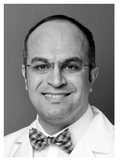
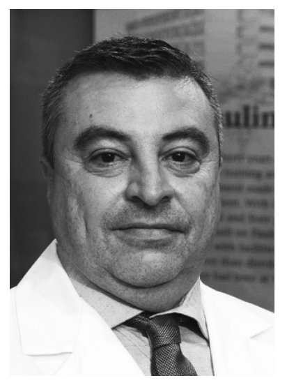
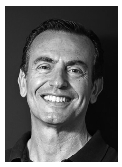
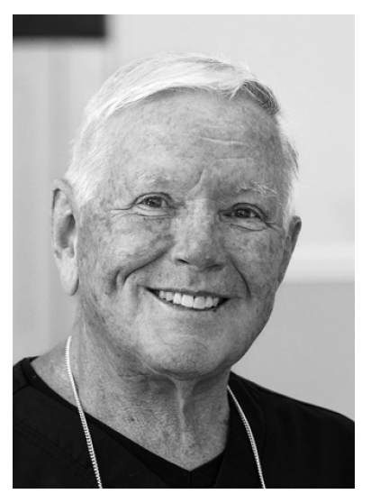
Drs. Elnagar and Sanchez are Associate Professors and Dr. Kusnoto is a Professor, Department of Orthodontics, College of Dentistry, University of Illinois Chicago, 801 S. Paulina St., Room 131, Chicago, IL 60612. Dr. Sambataro is an Adjunct Professor, Postgraduate Programs in Orthodontics, Department of Biomedical, Surgical and Dental Sciences, University of Milan “La Statale,” Milan, Italy. Dr. Sellke is in the private practice of orthodontics, Grayslake, IL. E-mail Dr. Elnagar at melnagar@uic.edu.