


CASE REPORT
Maxillary Expansion with the Invisalign Palatal Expander
Since transverse maxillary growth is the first dimension of craniofacial development to be completed, early intervention with rapid maxillary expansion (RME) is particularly advantageous in growing patients with narrow arches.1,2 By addressing transverse deficiencies prior to fusion of the midpalatal and circummaxillary sutures, RME can produce orthopedic effects to correct posterior crossbites, coordinate the maxillary and mandibular arches in Class II and III malocclusions, and increase arch perimeters for resolution of tooth-size/arch-size discrepancies.2
Previous short-term studies have shown that RME can successfully eliminate transverse discrepancies between the dental arches caused by maxillary constriction.3-5 Although few long-term RME studies have been conducted, several reports have indicated that such increases in the maxillary transverse dimension are relatively stable.6
Early approaches to RME relied on jackscrews and archwires.6 The literature on maxillary expansion using clear aligners has been limited, suggesting primarily dentoalveolar effects.6 The Invisalign* Palatal Expander (IPE) has recently been introduced as a viable alternative to screw and wire-based expansion devices, potentially inducing skeletal expansion in growing patients. Each sequential expander is printed from polyamide-12 (Nylon 12), eliminating the need for model fabrication.
Similar articles from the archive:
The IPE is .75mm thick at the buccal surfaces and 1.5mm at the occlusal surfaces, with a palatal thickness of 2.5-3mm.6 It contacts the lateral palatal walls but not the palatal vault. The tray covers the complete clinical crowns of the upper permanent molars and either the deciduous second molars or second premolars, plus half of the deciduous first molars or first premolars. Although lightweight, the expanders are rigid except at the buccal flanks, which have limited flexibility to enable removal using the built-in buccal handles.6
This article describes successful early maxillary expansion using the IPE in a growing patient.
Diagnosis and Treatment Plan
A 7-year-old female presented with minor asymmetry in the frontal view and a mildly excessive lower facial third (Fig. 1). The lateral profile was balanced, with a slightly open nasolabial angle, a normal labiomental sulcus, and lip competence. The patient had a Class I molar relationship on the right side and Class II molar and canine relationships on the left, along with normal overbite and overjet and a bilateral posterior crossbite.
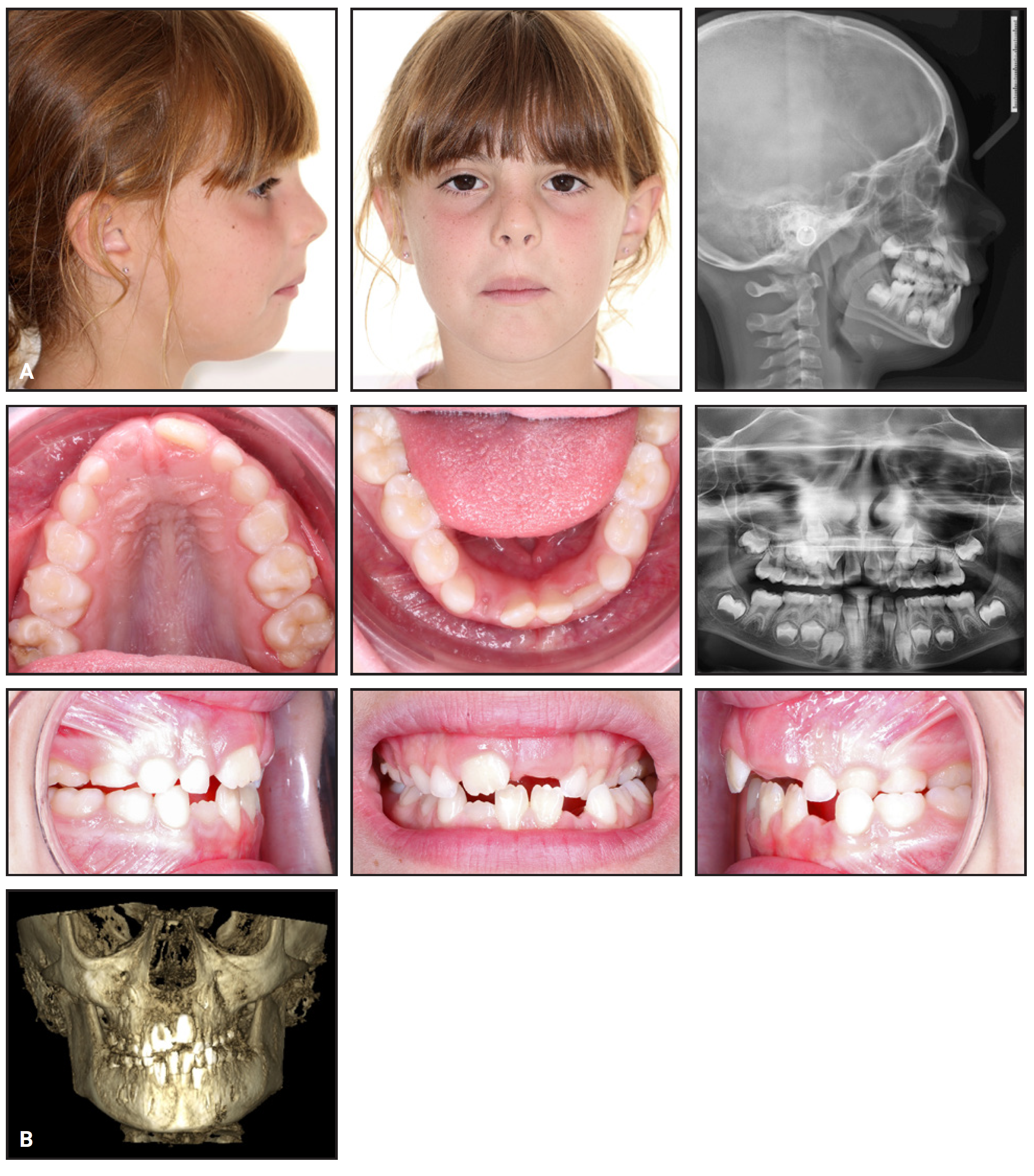
Fig. 1 A. 7-year-old female patient with maxillary transverse deficiency and bilateral posterior crossbite before treatment. B. Cone-beam computed tomography (CBCT) before treatment.
Cephalometric analysis found a normofacial pattern and a sagittal skeletal Class I relationship, accompanied by normal upper and lower incisor inclinations. TMJ evaluation, including conebeam computed tomography (CBCT), showed no signs or symptoms of dysfunction. These findings suggested normal craniofacial development, but indicated a transverse deficiency requiring maxillary expansion to maintain balanced growth and occlusion.
The primary treatment goals were to resolve the posterior crossbite, address the associated skeletal discrepancies, and create sufficient space for the erupting teeth. To accomplish these objectives, the maxillary arch would be expanded with the IPE. A ClinCheck* treatment plan is not generated for the IPE; instead, the prescription form specifies how much bilateral expansion is needed, from 0mm to 10mm. In this case, the prescription called for 10mm of expansion, and 40 expanders were ordered to achieve the planned correction, based on .25mm of expansion per day.
Treatment Progress
Attachments were bonded to the buccal surfaces
of the upper deciduous second molars and
permanent first molars to enhance retention and
to help maintain initial torque. The patient was
instructed to wear the IPE full-time, including
during meals, and to remove it only for regular oral
hygiene. She was asked to change the expander
daily, or every two days if sensitivity or poor fit
occurred. The first expander was passive to facilitate
adaptation (Fig. 2).
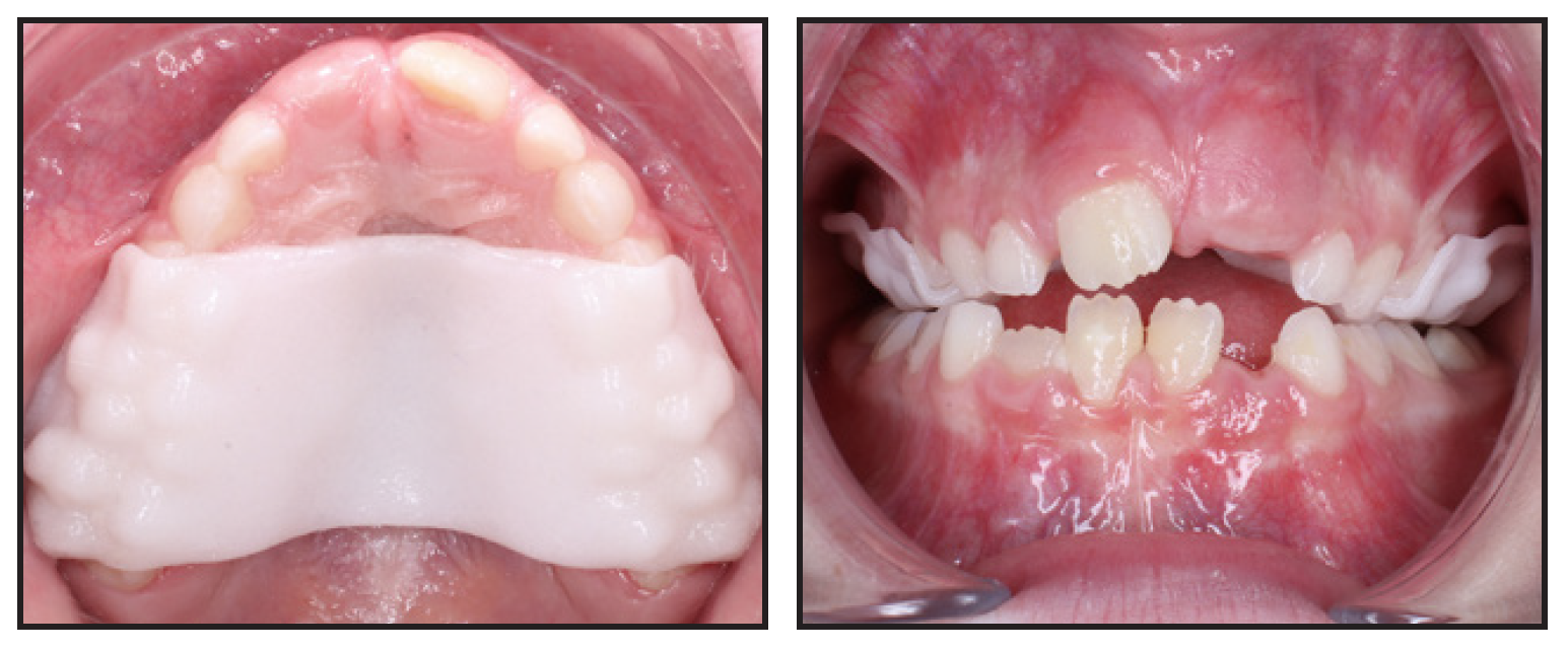
Fig. 2 First Invisalign* Palatal Expander (IPE) in place.
A follow-up visit was scheduled two weeks after the initial insertion to assess progress. The patient demonstrated excellent compliance; by changing the expanders daily, she had reached expander 14 at this point.
After 28 days of active expansion, the maxillary permanent first molars’ lingual cusps were contacting the mandibular molars, and therefore, active expansion was deemed complete after expander 28.
For the stabilization phase, 24 Invisalign Palatal Holders, replicating the final expansion stage, were delivered to be replaced weekly for six months.
Treatment Results
Total treatment duration was seven months. The patient maintained excellent oral hygiene and compliance throughout treatment. Her posterior crossbite was fully corrected; final records showed optimal esthetics and function, with no signs or symptoms of TMD (Fig. 3).
Post-treatment CBCT analysis demonstrated opening of the midpalatal suture, consistent with a skeletal component of expansion (Fig. 3B).
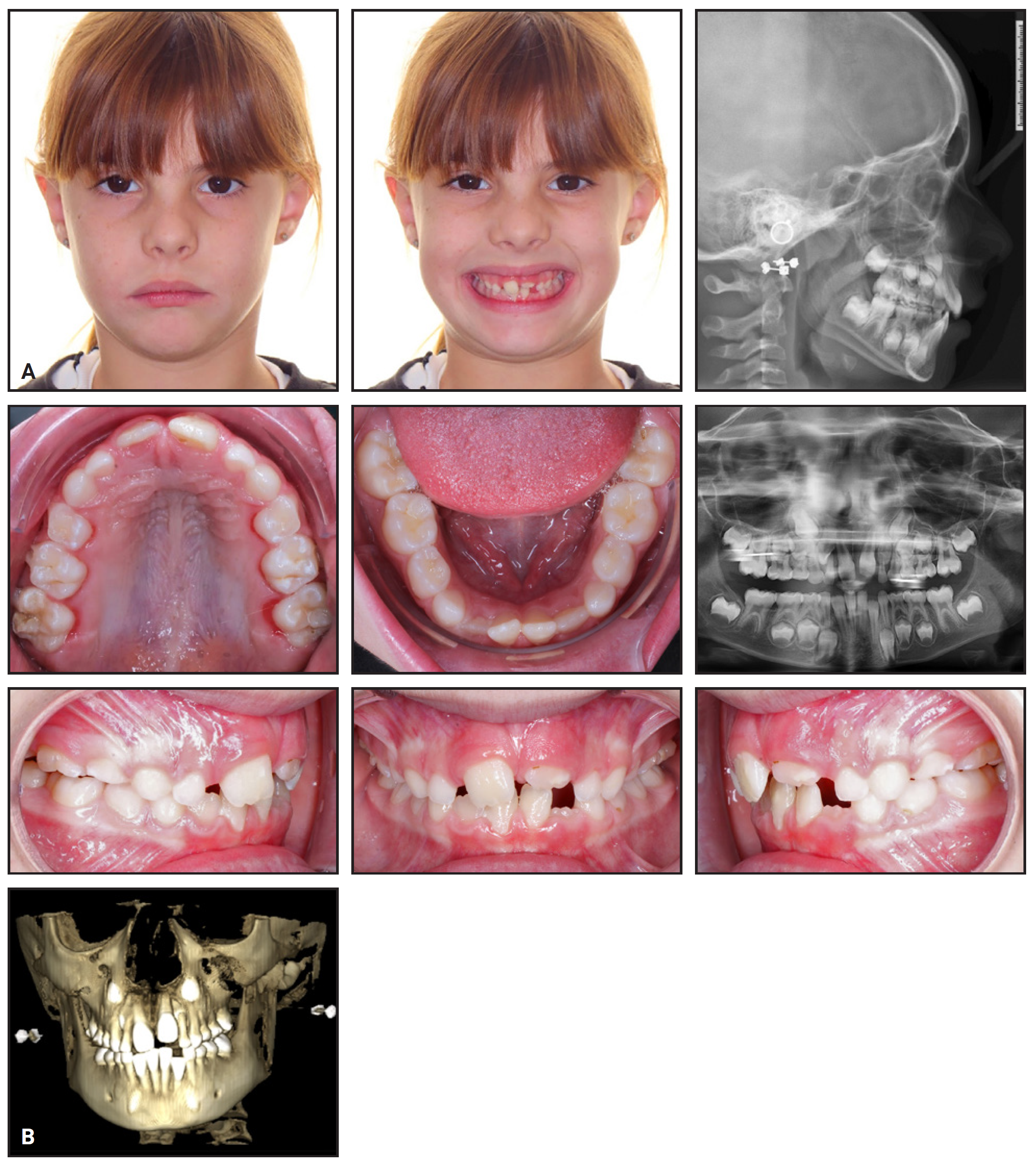
Fig. 3 A. Patient after seven months of treatment with IPE. B. CBCT after treatment.
Discussion
This case demonstrates the potential of the IPE as an effective and patient-friendly option for early orthodontic intervention. Treatment included attachments on the upper second deciduous molars and permanent first molars to optimize force distribution.
The patient was instructed to wear the IPE continuously, removing it only for brushing and flossing. She and her parent received comprehensive written and verbal instructions on the proper sequencing and use of the device. The patient quickly learned to insert and remove the expanders independently, and she reported that they felt comfortable. Weekly visits ensured close monitoring and timely adjustments when needed. Given the patient’s good compliance and oral hygiene, we observed no palatal or gingival inflammation, demineralization, or caries.
Although the treatment objectives were fully achieved, this case represents only one clinical experience. Further controlled studies are needed to confirm the efficacy and long-term stability of the IPE and to quantify the skeletal vs. dental components of expansion.
FOOTNOTES
- *Registered trademark of Align Technology, Inc., San Jose, CA; www.aligntech.com.
REFERENCES
- 1. Lee YS, Bayome M, Chou AHK, Vaiid N, Han SH, Kook YA. Transverse autocorrection of molar inclination in Class III patients: case Insights on maxillary protraction using Class III palatal plate. Semin Orthod. 2025;31(2):329-341. doi: https://www.doi.org/10.1053/j.sodo.2024.10.007
- 2. Walter A, Winsauer H, Crespo E, et al. Adult maxillary expansion: CBCT evaluation of skeletal changes and determining an efficiency factor between force-controlled polycyclic slow activation and continuous rapid activation for mini-screw-assisted palatal expansion—MASPE vs. MARPE. Head Face Med. 2024;20(1):70. doi: https://www.doi.org/10.1186/s13005-024-00474-6
- 3. Kravitz ND. Everybody gets an expander. Semin Orthod. 2023;29(2):171-173. doi: https://www.doi.org/10.1053/j.sodo.2023.05.002
- 4. Moshiri M, Kravitz ND, Nicozisis J, Miller S. Invisalign eighth-generation features for deep-bite correction and posterior arch expansion. Semin Orthod. 2021;27(3):175-178. doi: https://www.doi.org/10.1053/j.sodo.2021.09.002
- 5. Bouchant M, Saade A, El Helou M. Is maxillary arch expansion with Invisalign® efficient and predictable? A systematic review. Int Orthod. 2023;21(2):100750. doi: https://www.doi.org/10.1016/j.ortho.2023.100750
- 6. Nicozisis JL. Protocol for the Invisalign palatal expander. J Clin Orthod. 2024;58(12):725-730.
-
 DR. SABOUNI
 -
 DR. CARUSO
Dr. Sabouni is in private practice and an orthodontist consultant, United Arab Emirates, and a part-time lecturer, University of Dijon, France. Dr. Caruso is a professor, Department of Life, Health and Environmental Sciences, University of L’Aquila, Via Vetoio, 67100 L’Aquila, Italy; e-mail: silvia.caruso@univaq.it.