Favorite Saved

PEARLS

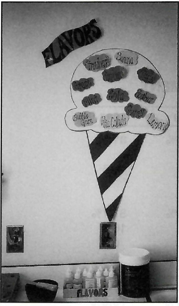
The alginate flavoring drops developed by various manufacturers have markedly improved young patients' reactions to taking impressions. We have designed a wall hanging that allows children to choose from the different flavors we have available. The "ice cream cone" is made from heavy poster board, and each flavor is backed with Velcro adhesive so it can be removed if it is temporarily out of supply.

Some young Phase I patients actually expect to be rewarded with ice cream after their impressions are taken.

Similar articles from the archive:

  • RICHARD L.
    DR. FOGEL
  • TAMYRA K.
    MS. BAILEY
  • JILL M.
    MS. DEEKS

Dr. Fogel in is Private Practice and Ms. Bailey and Deeks are Assistants at 84 E. Broad St. Elyria, OH 44035.

Fig. 1 Case 1. Pretreatment records.
Fig. 2 Cephalometric prediction tracing for mandibular advancement. A. Tracing of structures which will not be altered with surgery or orthodontics. B. Placement of distal mandible into optimum relation with remainder of skeleton. C. Addition of new A-Po line and corpus axis. D. Ideal placement of upper and lower incisors calculated position of molars and lips traced. E. Superimposed original and prediction tracing to evaluate overall treatment objectives and results.
Fig. 3 Case 2. Pretreatment records.
Fig. 4 Cephalometric prediction tracing for maxillary superior repositioning. A. Tracing of structures which will not be altered with surgery or orthodontics. B. Line parallel to the Frankfort horizontal at desired vertical level of maxillary central incisors. C. Autorotation of mandible around condyle as it will occur with the proposed amount of superior repositioning of the maxilla. D. Having added the proposed amount of genioplasty, a new A-Po line is drawn to the new Po as determined by the genioplasty. E. Placement of teeth as described.
Fig. 4a (Continued) F. Tracing of new nasal profile. G. Tracing of new upper lip position. H. Tracing of new lower lip position. I. Tracing of new soft-tissue chin. J. Superimposed original cephalometric tracing and prediction tracing.
Fig. 5 Cephalometric criteria which may be us d to help determine optimum anteroposterior soft-tissue chin position.
Fig. 6 Case 3. Pretreatment records.
Fig. 7 Cephalometric prediction tracing for combined maxillary and mandibular cases. A. Tracing of structures which will not be altered with surgery or orthodontics. B. Vertical referent parallel to Frankfort horizontal to determine new upper incisor position. C. Determination of anteroposterior position of maxilla and upper teeth. D. Placement of mandible into optimum position wlth maxilla. E. Finished prediction tracing. F. Superimposed original cephalometric tracing and prediction tracing.
Fig. 8 Utilizing the prediction tracing. A. Desired orthodontic change in the mandible. B. Desired orthodontic change in the maxilla. C. Desired position of the teeth prior to surgery to allow desired anteroposterior change at surgery. D. Superimposition showing desired orthodontic change following surgery.

DR. LEWARD C. FISH DDS, MS

DR. LEWARD C.  FISH DDS, MS
Orthodontist, Center for the Correction of Dentofacial Deformities, John Peter Smith Hospital, Fort Worth, Texas 76104.

DR. BRUCE N. EPKER DDS, PhD

DR. BRUCE N.  EPKER DDS, PhD
Director, Center for the Correction of Dentofacial Deformities, John Peter Smith Hospital, Fort Worth, Texas 76104.

My Account

This is currently not available. Please check back later.

Please contact heather@jco-online.com for any changes to your account.