


CASE REPORT
Hemisection of a Fused Maxillary Central Incisor with a Supernumerary Lateral Incisor in a Patient with Significant Maxillary Crowding
Fusion is described as the union of the enamel and/or dentin of two or more separate teeth.1-7 The prevalence of this tooth anomaly is estimated to be greater in the deciduous dentition (.5-2.5%) than in the permanent dentition (0.1-1.0%).8,9 Fused teeth can occur in either the mandible or the maxilla; in the permanent dentition, they are most commonly found in the maxillary anterior region.2,4 Although the etiology has yet to be determined, possible causes include trauma,2 environmental and genetic factors,1,4 and the presence of syndromes such as chondroectodermal dysplasia,3 achondrodysplasia, focal dermal hypoplasia, or osteopetrosis.8
Fusion can be partial or complete, depending on the stage of development at which the union occurred. The fusion is considered partial when the junction of two teeth includes only the crowns, and complete when there is a total union involving both crowns and roots.2,5 Multidisciplinary therapeutic options include hemisection of the fused counterpart, root canal treatment, and restoration with composite or a full crown.1,3,5,9
This case report describes the orthodontic management of a patient with a fused upper left central and supernumerary lateral incisor, a lingually displaced lateral incisor, and bilaterally impacted upper canines.
Diagnosis and Treatment Plan
An 11-year-old female presented with crowding in both arches and complete fusion of the upper left central incisor to a supernumerary lateral incisor. There was no significant medical history and no family history of any dental anomalies. Clinical examination showed a mesofacial, symmetrical face with balanced facial thirds and a slightly convex profile (Fig. 1).
Similar articles from the archive:
- CASE REPORT Hemisection of an Ankylosed Lower Second Deciduous Molar for Orthodontic Closure of a Congenitally Missing Second-Premolar Space June 2024
- Controlled Slicing for Space Closure in Cases with Congenitally Missing Second Premolars April 2023
- CASE REPORT Translation of a Tooth Across the Suture to Replace a Fused Upper Central Incisor June 2022
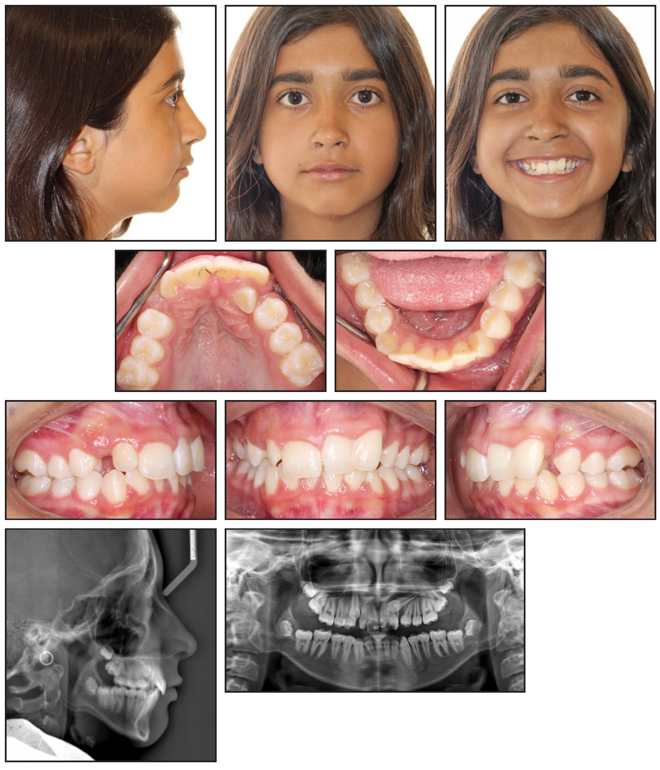
Fig. 1 11-year-old female with fused upper left central and supernumerary lateral incisor, lingually displaced upper left lateral incisor, bilaterally impacted upper canines, and significant maxillary crowding before treatment.
The fused incisors were similar in size, with a combined mesiodistal width approximating that of the added width of the contralateral lateral and central incisors. In addition, the patient exhibited a lingually displaced upper left lateral incisor, a horizontally impacted upper left canine, a vertically impacted upper right canine, and a lingual wire splint between the central incisors. A gingival cleft was observed on the labial aspect of the alveolar ridge between the supernumerary lateral incisor and the upper left first premolar. The patient had a slightly excessive upper right incisor display in smiling, as well as an increased overbite with a cant in both central incisors. There was a slight Class II molar relationship on the right side and an end-on Class II relationship on the left side, with significant maxillary crowding and mild mandibular crowding. Both maxillary and mandibular dental midlines coincided with the facial midline.
Radiographic images and cone-beam computed tomography (CBCT) scans revealed a cleft where the crown of the upper left central incisor was fused with the supernumerary lateral incisor, including the cervical third of the root (Fig. 2). The upper left lateral incisor was positioned directly lingual to the fused central incisor, and the crown of the impacted upper left canine was lingual and apical to the lateral incisor. The maxillary left buccal segment had collapsed lingually, creating a crossbite tendency in the first premolar region.
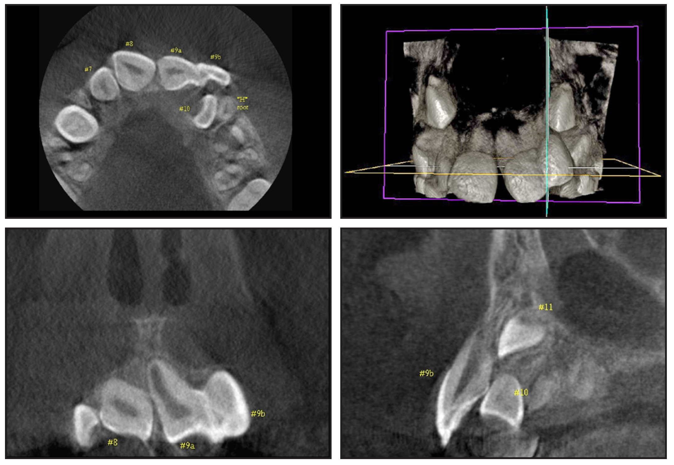
Fig. 2 Cone-beam computed tomography scans showing completely fused upper left central and supernumerary lateral incisor, as well as palatally impacted upper left canine.
The proposed treatment involved hemisection of the completely fused upper left central incisor and supernumerary lateral incisor. Extraction of the supernumerary would be preceded by root canal therapy, using calcium hydroxide to obturate the lateral canals communicating with the pulp of the central incisor. Vitality of the remaining central incisor would be preserved by avoiding root canal therapy. Extraction of the upper left first premolar was also planned to allow labial positioning of the lateral incisor and impacted canine. The maxillary and mandibular crowding would be relieved orthodontically, in conjunction with reshaping, recontouring, and restoration of the upper left central incisor.
Another potential treatment option involved extraction of the fused teeth, with the palatally impacted upper left lateral incisor substituting for the central incisor and the canine substituting for the lateral incisor, but this would have created large tooth-size asymmetries in the esthetic zone. Orthodontic treatment could also have been performed without sectioning the fused tooth, maintaining the lateral incisor in its original lingual position while distalizing the left buccal segment or extracting the first premolar to allow eruption of the impacted canine. When the patient reached adulthood, the fused tooth would be extracted, an implant would immediately be placed in the central incisor site, and the lingually displaced lateral incisor would be orthodontically repositioned labially. Considering all the potential risks and benefits, however, the parents and patient chose hemisection of the fused teeth.
Treatment Progress
MBT preadjusted .022" brackets were bonded in the maxillary arch, bypassing the upper left first premolar, to achieve leveling and alignment of the upper central incisors with slight extrusion of the fused teeth. Seven months later, the patient was referred to a periodontist for extraction of the upper left first premolar, hemisection of the fused teeth, and exposure of the impacted canine. Before extraction of the fused supernumerary lateral incisor, root canal therapy was performed with calcium hydroxide to ensure obturation of the lateral canals leading to the fused central incisor’s root. The central incisor was then sectioned from the fused supernumerary and reshaped to resemble the proper contour of a central incisor. The periodontist used a window technique to expose the impacted canine, rather than raising a flap in the thick palatal tissue, to avoid increasing the mobility of the lateral incisor. The cortical bone of the palate was removed to expose the canine crown, and a gold chain was bonded to the lingual surface of the tooth to continue with orthodontic treatment.
The lingually displaced upper left lateral incisor moved labially to the desired position without any applied force (Fig. 3). MBT preadjusted .022" brackets were then bonded in the mandibular arch. The maxillary and mandibular crowding were corrected, and the desired occlusion was achieved with elastics (3/16", 6oz).
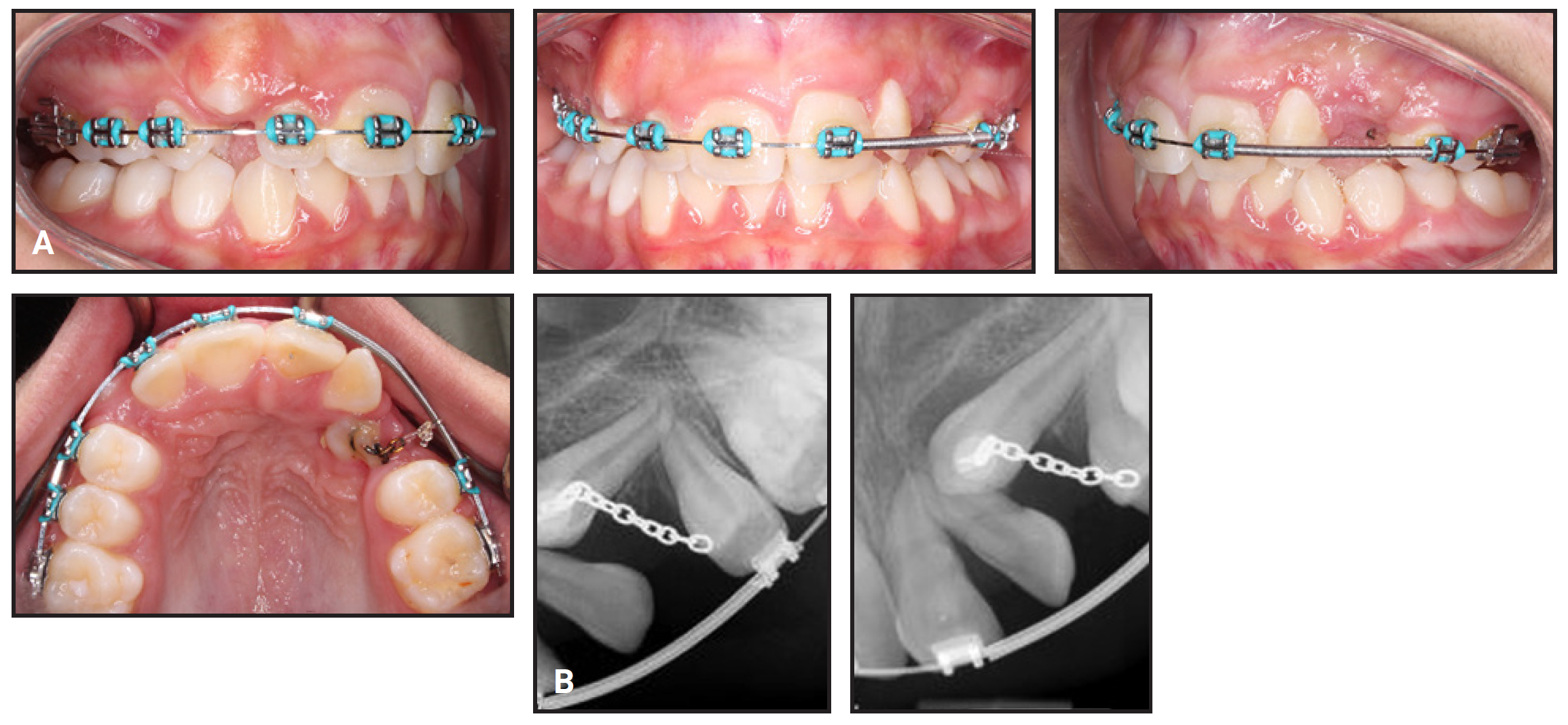
Fig. 3 A. After seven months of treatment, showing leveling and alignment of maxillary arch and self-correction of lingually displaced upper left lateral incisor. B. Periapical radiographs of impacted upper left canine after bonding of gold chain for traction.
During the finishing phase, we noted that the root of the central incisor was abnormally tipped distally, which precluded a perfect match of its incisal edge to that of the contralateral incisor (Fig. 4). Because the lower second molars were also impacted and tipped distally, the third molars were extracted.
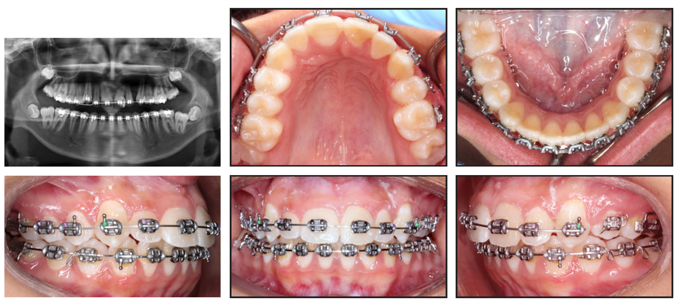
Fig. 4 After 22 months of treatment, panoramic radiograph showing abnormal distal root tip of upper left central incisor and bilaterally impacted and distally tipped lower second molars.
Treatment Results
The vitality of the hemisected upper left central incisor was maintained throughout treatment. The impacted left canine was erupted into its proper position, although a vertical alveolar bone deficiency persisted over the long term (Fig. 5).
After 58 months of orthodontic treatment, a slight recession of the upper left canine was noted, but the overall smile esthetics and occlusal results were good (Fig. 6). A restorative dentist built up the distal aspect of the upper left central incisor with composite. Although a veneer was planned for the future, the patient is currently satisfied with her smile esthetics.
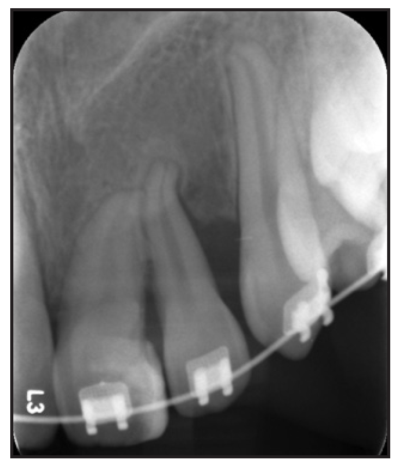
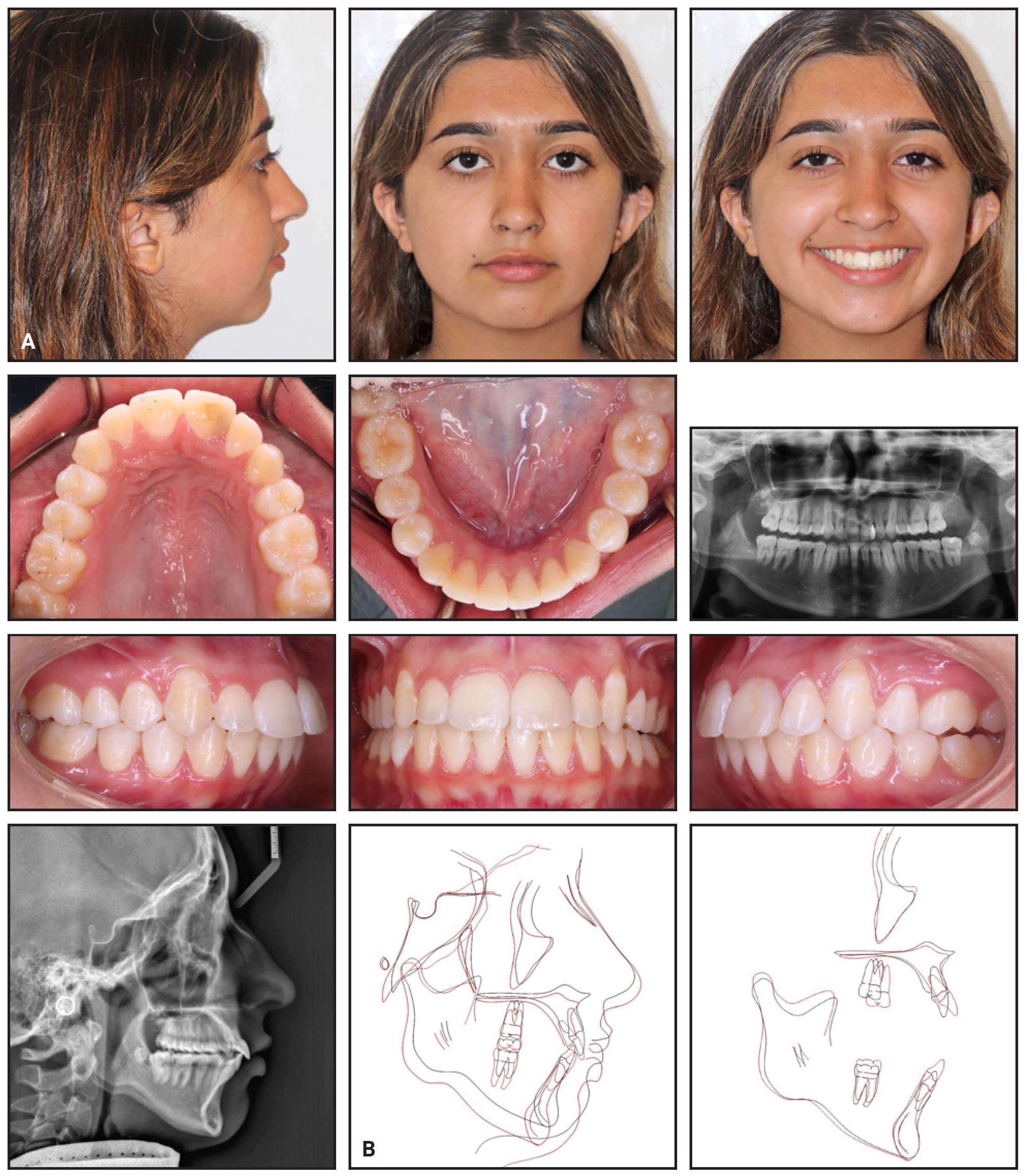
Fig. 6 A. Patient after 58 months of treatment. B. Superimposition of pretreatment (black) and post treatment (red) cephalometric tracings.
Three years after debonding, the results remained stable (Fig. 7). The labial gingival margin of the upper left canine had crept coronally, showing no signs of long-term recession. Vacuum-formed retainers were prescribed to be worn for 16 hours per day for the first six months, followed by nighttime wear thereafter. Hawley retainers were also delivered, to be alternated with the vacuum-formed retainers on a daily basis during nighttime wear.
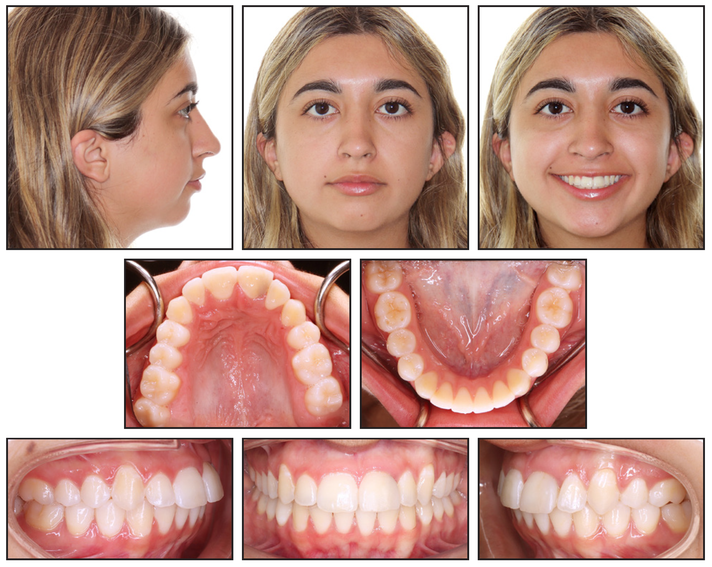
Fig. 7 Patient three years after treatment.
Discussion
Dental anomalies such as fusion can manifest in various ways. If the fusion occurs between two normal teeth, there might be spacing or a diastema along the arch. On the other hand, if the fusion involves a supernumerary tooth, some degree of crowding may be present. Either scenario can result in malocclusion, malpositioning, and severe esthetic problems causing psychological complications, particularly in children and adolescents.9
Treatment of tooth fusion will differ according to the severity and complexity of the case and factors such as the location of the connecting area and the patient’s age and developmental stage when the junction occurred.8 A conservative protocol might involve mesiodistal interproximal reduction and recontouring, followed by prosthodontic restoration with composite or a full-coverage crown to mimic the shape of the appropriate tooth.4 Other treatment approaches include extraction, ex vivo separation of the tooth followed by reimplantation into its original location,8 separation into two single teeth, dissection and removal of the nonessential portion after root canal treatment, and autotransplantation after extraction.9 In-depth radiographic analysis and consultation with multiple specialists are recommended to establish a comprehensive treatment plan.
In the case shown here, after evaluating the CBCT and clinically inspecting the area during extraction of the upper left first premolar, the periodontist confirmed that it was possible to section and remove the supernumerary portion of the central incisor, creating a far more symmetrical appearance of the remaining tooth. Our patient had the added advantage of a thick gingival biotype. The cleft where the central incisor was fused with its supernumerary counterpart was deep and widened at the coronal aspect, creating a pseudo-papilla that would ideally develop into a true papilla with a proper col once the lateral incisor was brought into the arch.
Multiple reports have shown that vitality of the remaining tooth segment can be maintained after a hemisection.1,3,5,8,9 Previous case reports have shown that root canal therapy is not always needed because fused teeth tend to have two separate roots and root canal systems.1,3,8 Hülsmann and colleagues are the only authors to report performing a root canal on the segment that was going to be extracted.5 After evaluation of the pulp cavity of the mesial part of the fused tooth, they cleaned, shaped, temporized with calcium hydroxide, and sealed the tooth cavity. The patient reported no changes or symptoms, and the distal part of the tooth remained vital one week later.
In our patient, the fusion between the central incisor and the supernumerary lateral incisor occurred in the coronal portion of the root trunk and also had two separate roots and canal systems, but with a small lateral canal directly connecting the two segments. After detailed evaluation of the CBCT scan, the endodontist decided to clean, shape, and seal the open lateral canal with calcium hydroxide to promote and maintain the vitality of the central incisor. No root canal treatment was performed on the central incisor because the endodontist found no visible communication between the two segments at the coronal level, in a microscopic evaluation after accessing the pulp chamber of the supernumerary tooth.
Following hemisection and removal of the supernumerary portion of the fused tooth, the remaining segment was restored using composite resin. Although several case reports have mentioned postoperative side effects such as hypersensitivity, external root resorption, and reversible pulpitis,8 no radiographic or clinical pathological findings were noted in our patient. The remaining tooth segment responded normally to pulp-vitality tests, and long-term monitoring showed the multidisciplinary treatment to be effective in reestablishing both function and esthetics. A slight shade contrast was noticeable between the distal composite restoration and the hemisected tooth. A veneer was proposed to the patient to mask this discrepancy, but she postponed consideration of this more extensive restoration.
In the cephalometric analysis, a slight protrusion of the maxillary incisors and the mandibular dentition was noted at the end of treatment (Table 1).
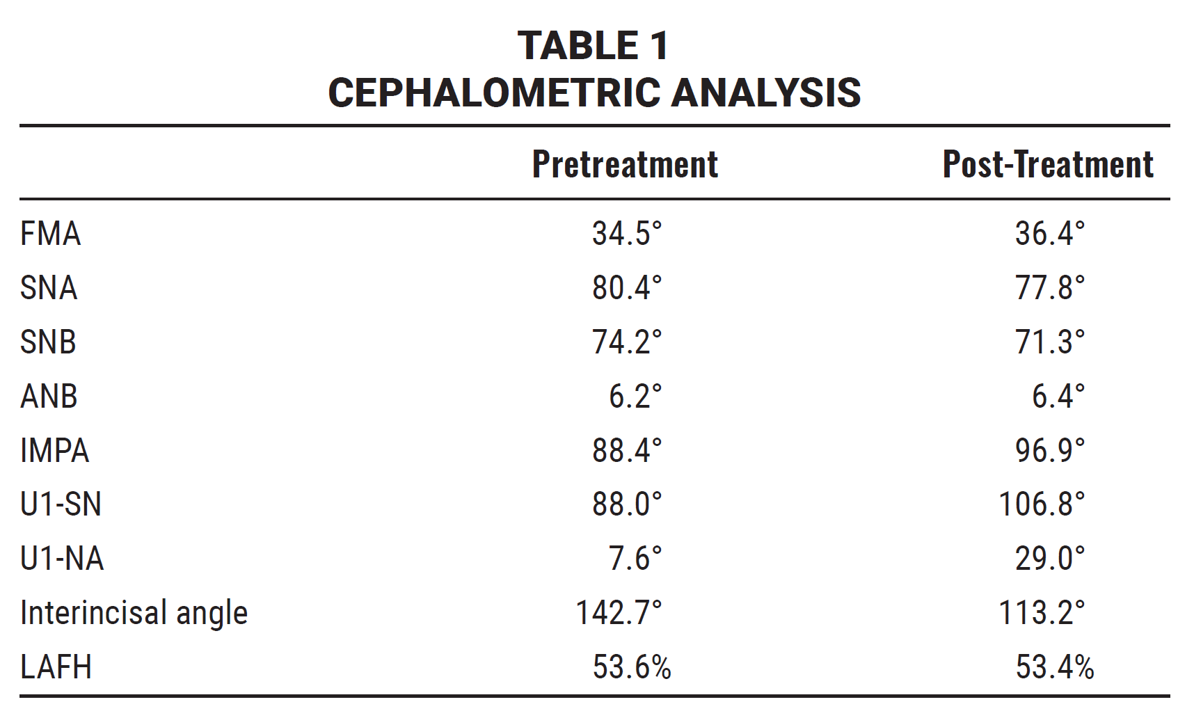
The upper left first premolar was extracted because of excessive crowding in the maxillary left quadrant. Since the maxillary incisors were slightly upright before treatment, and a minor correction of their inclination was needed, we elected not to extract a tooth in the maxillary right quadrant. That decision was further supported by the lip relationship, which showed an adequate anteroposterior position with no lip strain or interlabial gap. This lip position was maintained throughout treatment, as shown in the cephalometric superimpositions (Fig. 6B).
From a biomechanical perspective, further reduction of the incisor inclination would have required extraction of the other three first premolars, with skeletal anchorage needed for distalization of the maxillary left quadrant. Bilateral distalization of the maxillary posterior segments, in conjunction with the upper left premolar extraction and interproximal reduction in the mandibular arch, was another possibility. Both approaches would have required temporary anchorage devices, however, which would have significantly increased the treatment time in this already complex case, while increasing biomechanical demands and costs.
REFERENCES
- 1. Cetinbas T, Halil S, Akcam MO, Sari S, Cetiner S. Hemisection of a fused tooth. Oral Surg Oral Med Oral Pathol Oral Radiol Endod. 2007;104(4):e120-e124. doi: https://www.doi.org/10.1016/j.tripleo.2007.03.029
- 2. Chen F, Wang S, Bai N, et al. Management of fused anterior teeth: review and clinical report. J Prosthodont. 2022;31(4):282-288. doi: https://www.doi.org/10.1111/jopr.13478
- 3. Finkelstein T, Shapira Y, Bechor N, Shpack N. Surgical and orthodontic treatment of a fused maxillary central incisor and supernumerary tooth. J Clin Orthod. 2014;48(10):654-658.
- 4. Finkelstein T, Shapira Y, Bechor N, Shpack N. Fused and geminated permanent maxillary central incisors: prevalence, treatment options, and outcome in orthodontic patients. J Dent Child (Chic). 2015;82(3):147-152.
- 5. Hülsmann M, Bahr R, Grohmann U. Hemisection and vital treatment of a fused tooth—literature review and case report. Endod Dent Traumatol. 1997;13(6):253-258. doi: https://www.doi.org/10.1111/j.1600-9657.1997.tb00051.x
- 6. Over LM, Sadowsky SJ, LeMert S, Dryden M. Interdisciplinary treatment for an adolescent with fusion of the left maxillary lateral and central incisors: a clinical report. J Prosthet Dent. 2025;134(4):875-878. doi: https://www.doi.org/10.1016/j.prosdent.2023.11.013
- 7. Worthy CE, Wilson OL, Hudson JW. Concurrence of gemination and fusion in maxillary central incisors: a case report. J Tenn Dent Assoc. 2016;96(1):27-31.
- 8. Demircioglu Guler D, Sen Tunc E, Arici N, Ozkan N. Multidisciplinary management of a fused tooth: a case report. Case Rep Dent. 2013;2013:634052. doi: https://www.doi.org/10.1155/2013/634052
- 9. Kim SY, Choi SC, Chung YJ. Management of the fused permanent upper lateral incisor: a case report. Oral Surg Oral Med Oral Pathol Oral Radiol Endod. 2011;111(5):649-652. doi: https://www.doi.org/10.1016/j.tripleo.2010.11.015
-
 DR. MONTES
-
 DR. ABEDI
-
 DR. URIBE
Dr. Luna is in private practice, 29320 Royal Aberdeen, Lake Elsinore, CA 92530; e-mail: marialunadds@outlook.com. Dr. Abedi is an endodontist in private practice, Middletown, CT. Dr. Uribe is a Contributing Editor of the Journal of Clinical Orthodontics and the Ravi Nanda Professor and Chair, Department of Orthodontics, University of Connecticut School of Dental Medicine.