



Continuing Education from June 2025 Issue (questions only)
Case #9, Part 2: Treatment
In this monthly column, Contributing Editors Gayle Glenn, DDS, MSD, and Shawn L. Miller, DMD, MMSc, present a case to challenge your diagnosis and treatment-planning skills. The initial records for this case were introduced in the previous issue. Here, one of the editors discusses how the case was treated.
Similar articles from the archive:
Diagnosis
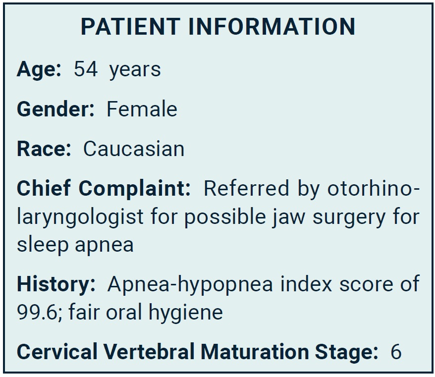
This patient was referred by her otolaryngologist for orthodontic treatment and orthognathic surgery to address her obstructive sleep apnea (OSA), as indicated by an apnea-hypopnea index (AHI) score of 99.6.
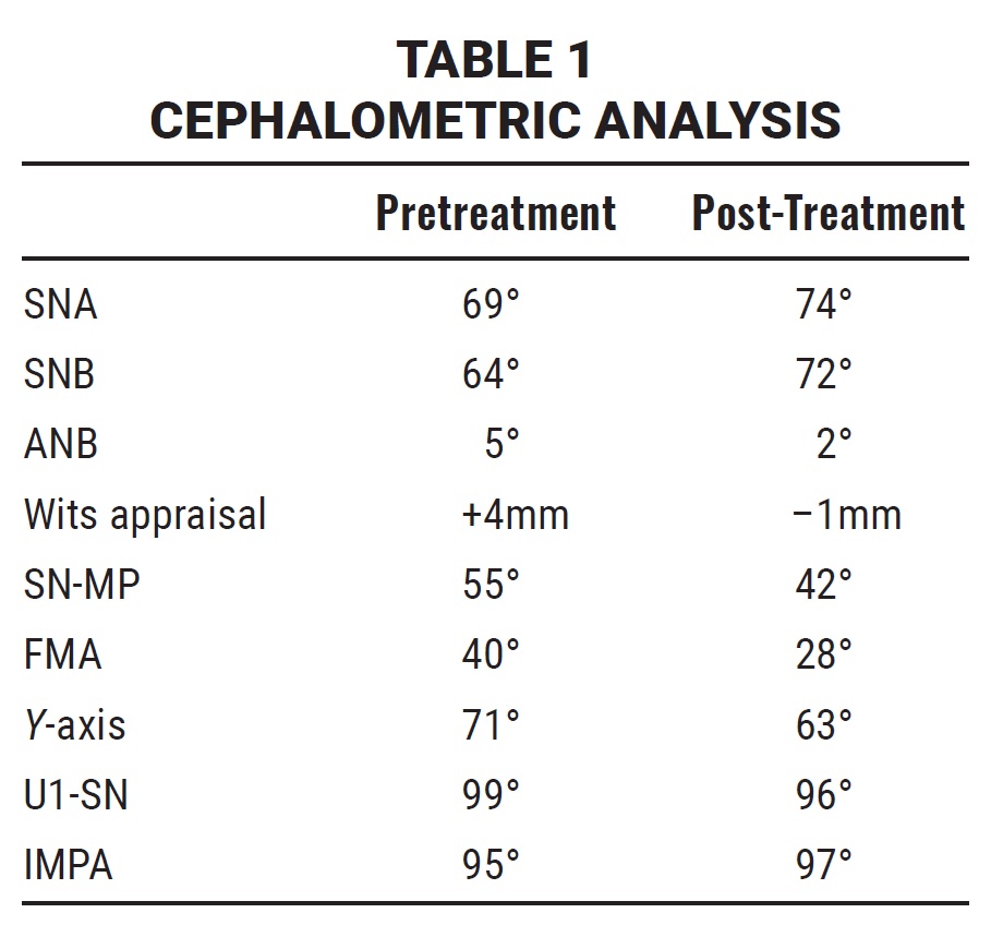
The patient was diagnosed with a skeletal Class II malocclusion. Clinical examination found a retrognathic maxilla and mandible, a hyperdivergent growth pattern, and increased lower anterior facial height—all craniofacial features associated with OSA. The maxillary and mandibular midlines were both deviated to the right of the facial midline. In smiling, a reverse smile arc was noted, along with wide buccal corridors and excessive gingival display in both the anterior and posterior regions.
End-on Class II molar and canine relationships were present bilaterally. The maxillary and mandibular archforms were both tapered, with moderate upper crowding and severe lower crowding. An anterior open bite was observed, along with an anterior crossbite at the upper right lateral incisor and a posterior crossbite at the upper right first premolar. The upper incisors were proclined, and the overjet was excessive. The upper lateral incisors were small, indicating a tooth-size discrepancy (Fig. 1A and 1B).
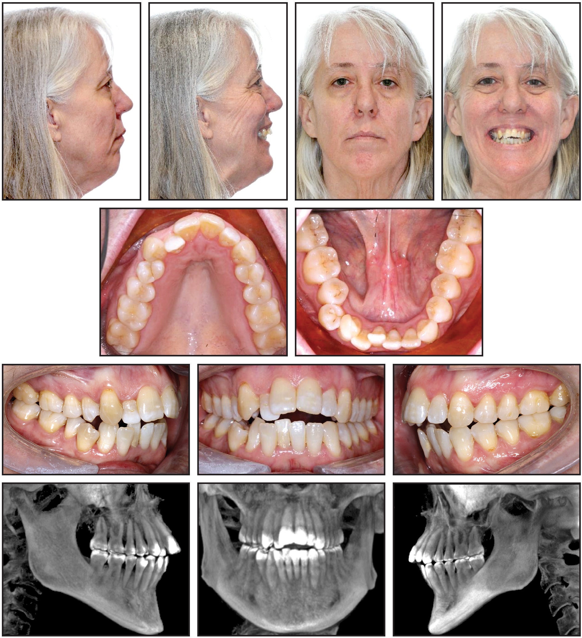
Fig. 1 Initial records (continued in next image).
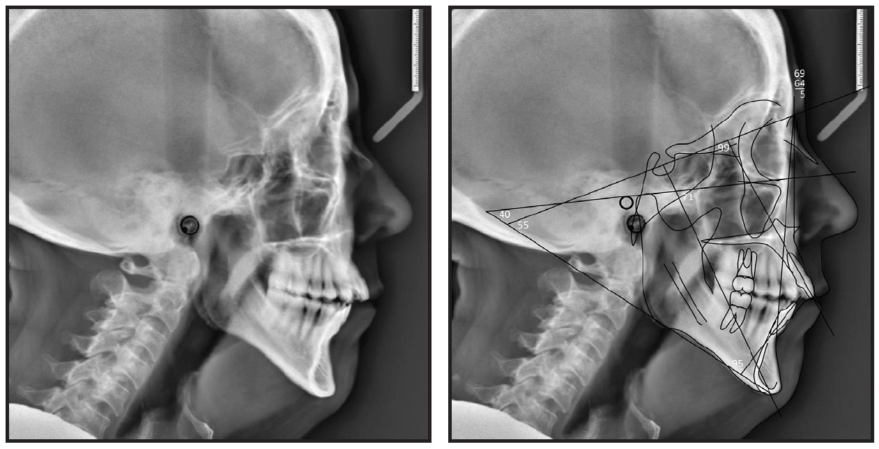
Fig. 1 (cont.) Initial records.
Discussion
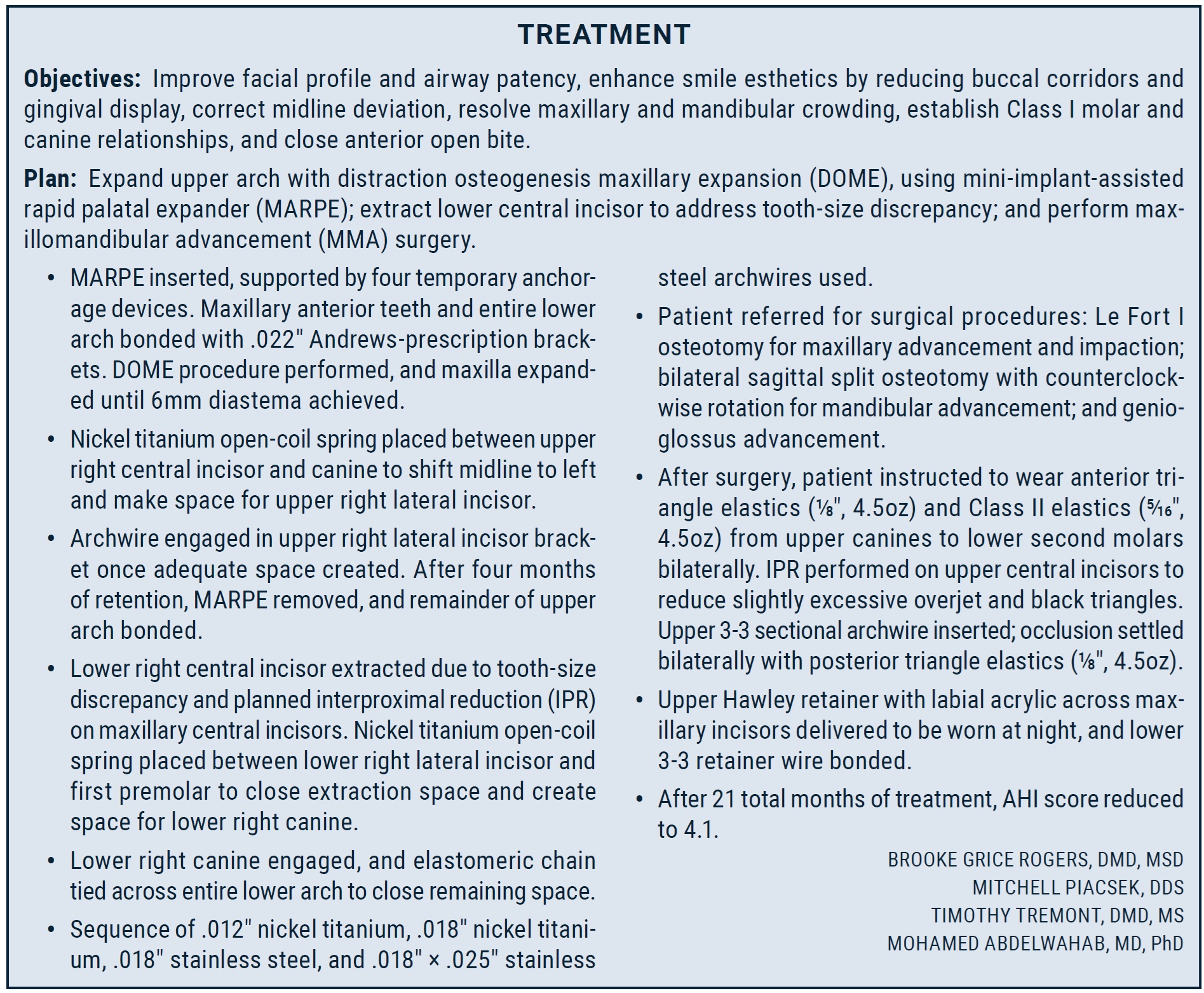
Over the past three decades, the use of orthodontics in conjunction with sleep surgery to treat adults with OSA has increased. This case highlights the importance of communication and coordination among the physician, the orthodontist, and the sleep surgeon to ensure a successful outcome. The notable reduction in AHI score, from 99.6 to 4.1, is likely to significantly enhance this patient’s overall health and wellness.
The patient was treated with two separate extrapharyngeal procedures—DOME for correction of the maxillary skeletal/dental transverse deficiency and nasal floor expansion, and MMA surgery to increase the total pharyngeal airway. MMA surgery is reported to have a high success rate and moderate cure rate, with the majority of patients expressing satisfaction with their postsurgical appearance.
In this case, the patient benefited from skillful planning and execution of both the surgical procedures and the orthodontic treatment. The DOME procedure provided significant maxillary expansion, allowing for a subsequent one-piece Le Fort I maxillary advancement and impaction. Extraction of a mandibular incisor addressed the mandibular crowding and the tooth-size discrepancy created by the small maxillary lateral incisors, enabling more mandibular advancement than if the patient were treated without extractions. Counterclockwise rotation of the maxillomandibular complex and a genioglossus advancement to protract the tongue base contributed to the enhanced hypopharyngeal airway and improved soft-tissue outcome. Together with IPR of the maxillary central incisors, this course of treatment resulted in a functional occlusion and esthetic improvement (Fig. 2A and 2B).
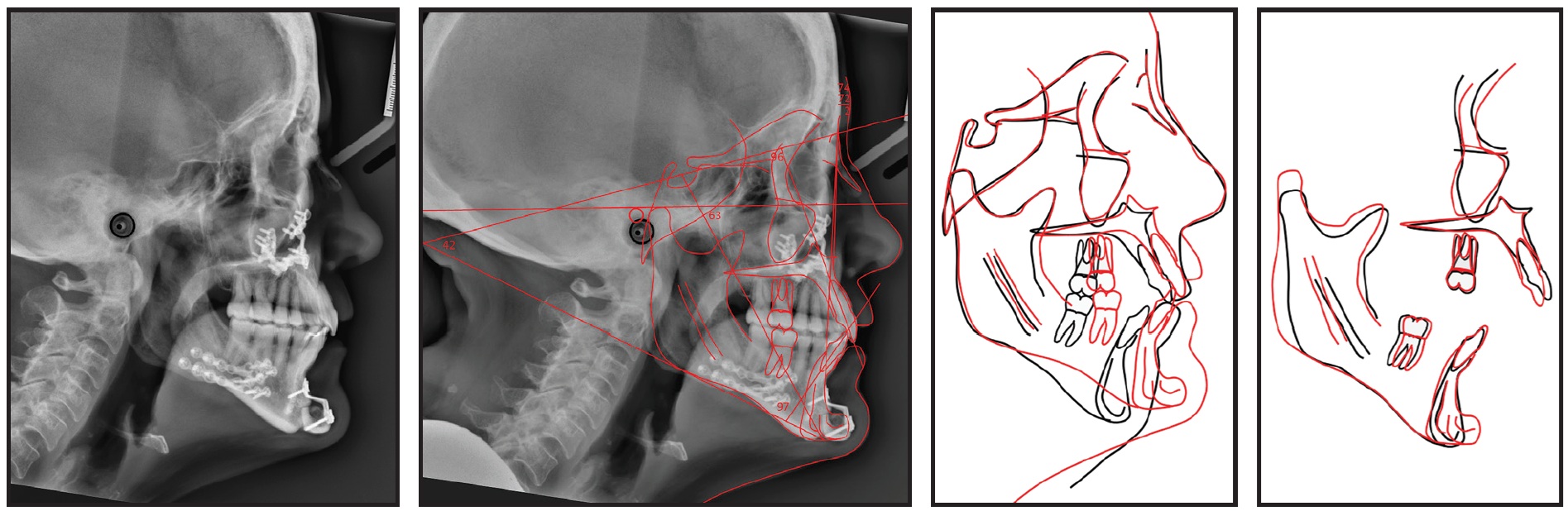
Fig. 2 Final records (continued in next image).
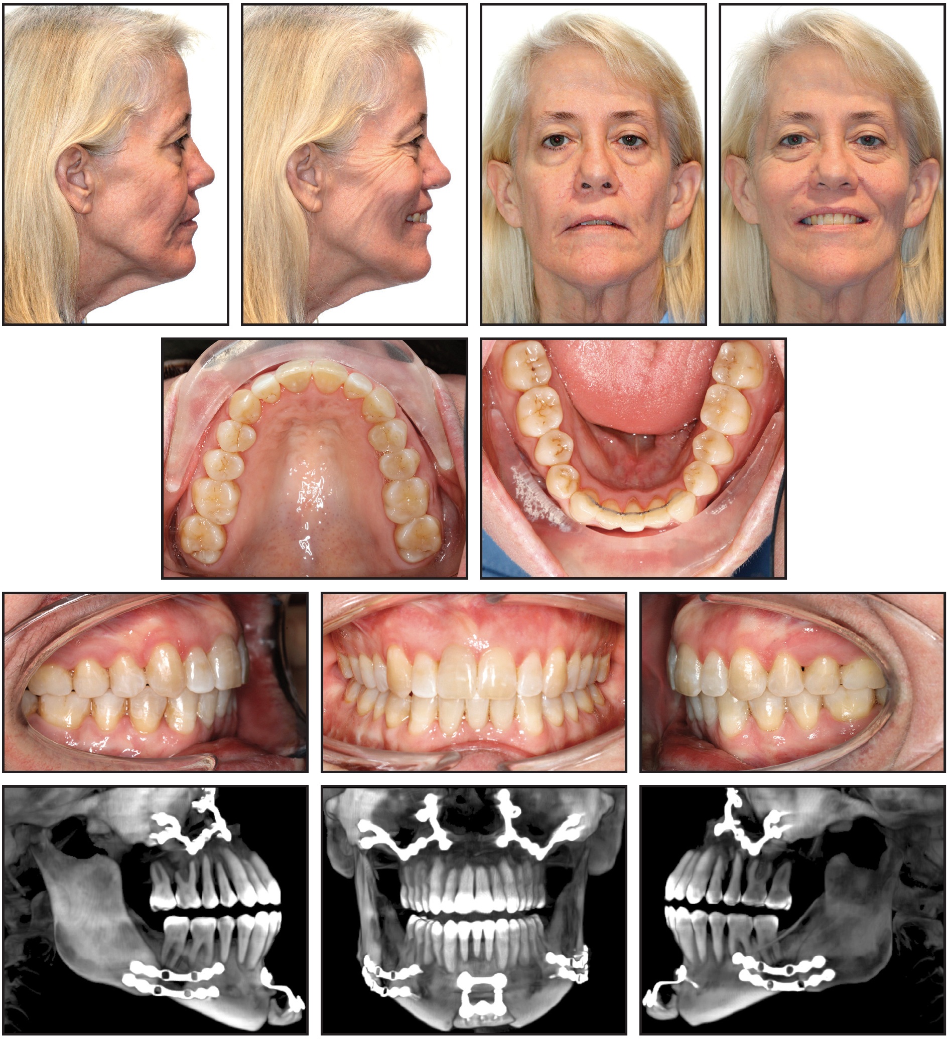
Fig. 2 (cont.) Final records.
A two-stage surgical approach was used, with DOME performed first to address the airway concerns, as well as the maxillary arch constriction, crowding, and incisor proclination. It is unknown whether a similar reduction in AHI score could have been achieved with a multipiece maxillary osteotomy performed in conjunction with the MMA surgery and genioglossus advancement.
GG
-
 DR. ROGERS
-
 DR. PIACSEK
-
 DR. TREMONT
-
 DR. ABDELWAHAB
-
 DR. GLENN
Dr. Rogers was an Orthodontic Resident, Dr. Piacsek is an Orthodontic Resident, Dr. Tremont is Professor and Head of Orthodontics, and Dr. Abdelwahab is Assistant Professor, Medical University of South Carolina, 173 Ashely Ave., BSB 347, MSC 507, Charleston, SC 29425. Dr. Rogers is now in Private Practice in Charleston, SC. E-mail Dr. Tremont at tremont@musc.edu. Dr. Glenn is a Contributing Editor of the Journal of Clinical Orthodontics.
