CASE REPORT
In-House 3D-Printed Shape Memory Aligners for Retreatment after Fixed Retainer Failure
Recent advances in materials science1 and computer-aided design and manufacturing (CAD/CAM) technology,2 in combination with the growing number of adult patients seeking inconspicuous orthodontic treatment,3 have led to the widespread adoption of clear aligners as a comprehensive alternative to fixed appliances.4 Increasingly, the digital design and fabrication of clear aligners are occurring “in-house”—within a private practice.5 The introduction of the first resin specifically intended for clear aligners6 now allows trays to be 3D-printed with greater dimensional precision than thermoformed aligners,7 while incorporating features such as planned variations in thickness across the aligner body and thermomechanical shape memory.6,8,9
To date, the literature on 3D-printed aligners largely consists of material6,10,11 and in vitro studies,9,12,13 with few investigations of their clinical efficacy. This article describes the digital design and in-house fabrication of customized aligners with shape memory to treat a case of failed fixed-wire retention, preventing the loss of a lower lateral incisor.
Diagnosis and Treatment Plan
A 32-year-old female who had previously undergone successful orthodontic treatment elsewhere presented to our practice with the chief complaint of a protrusive lower left canine (Fig. 1). Multistranded-wire lingual retainers had been bonded to the upper and lower incisors and canines. One year prior to our consultation, the retainer wire had come loose from the lower left canine, but it had been reattached before the patient noticed any changes in tooth position. The patient reported that within six months of rebonding, however, the lower left canine had moved significantly.
Clinical examination found a bilateral Class I occlusion with an open bite in the region of the lower left canine cusp, resulting in an asymmetrical lower arch due to the protrusion of the lower left incisors and proclination of the lower left canine. This proclination had caused the contours of the canine root to become visible on the lingual side. Gingival recession of 5mm, with no apparent inflammation, was noted on the buccal side of the lower left lateral incisor.
Similar articles from the archive:
- ALIGNER CORNER EX 30 vs. SmartTrack Materials in Maxillary Expansion with the Invisalign System June 2022
- ALIGNER CORNER Optical Properties of Seven Types of Clear Aligners Before and After In Vitro Aging March 2022
- ALIGNER CORNER Integration of In-House Aligner Therapy in Private Practice August 2021
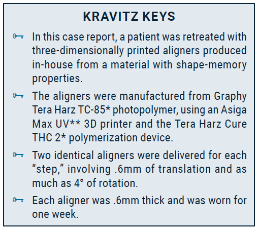
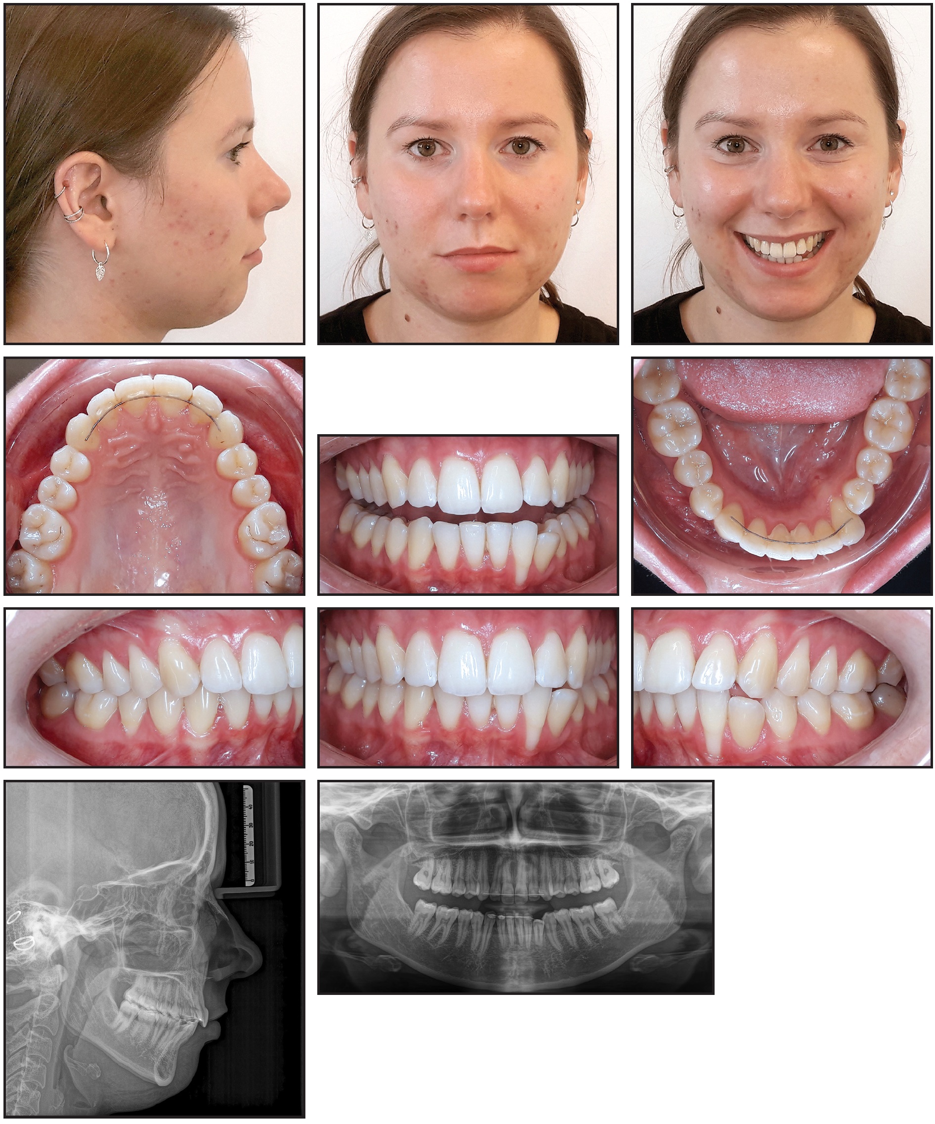
Fig. 1 32-year-old female patient with severe protrusion of lower left central incisor, lateral incisor, and canine due to failure of fixed lingual retainer, before retreatment.
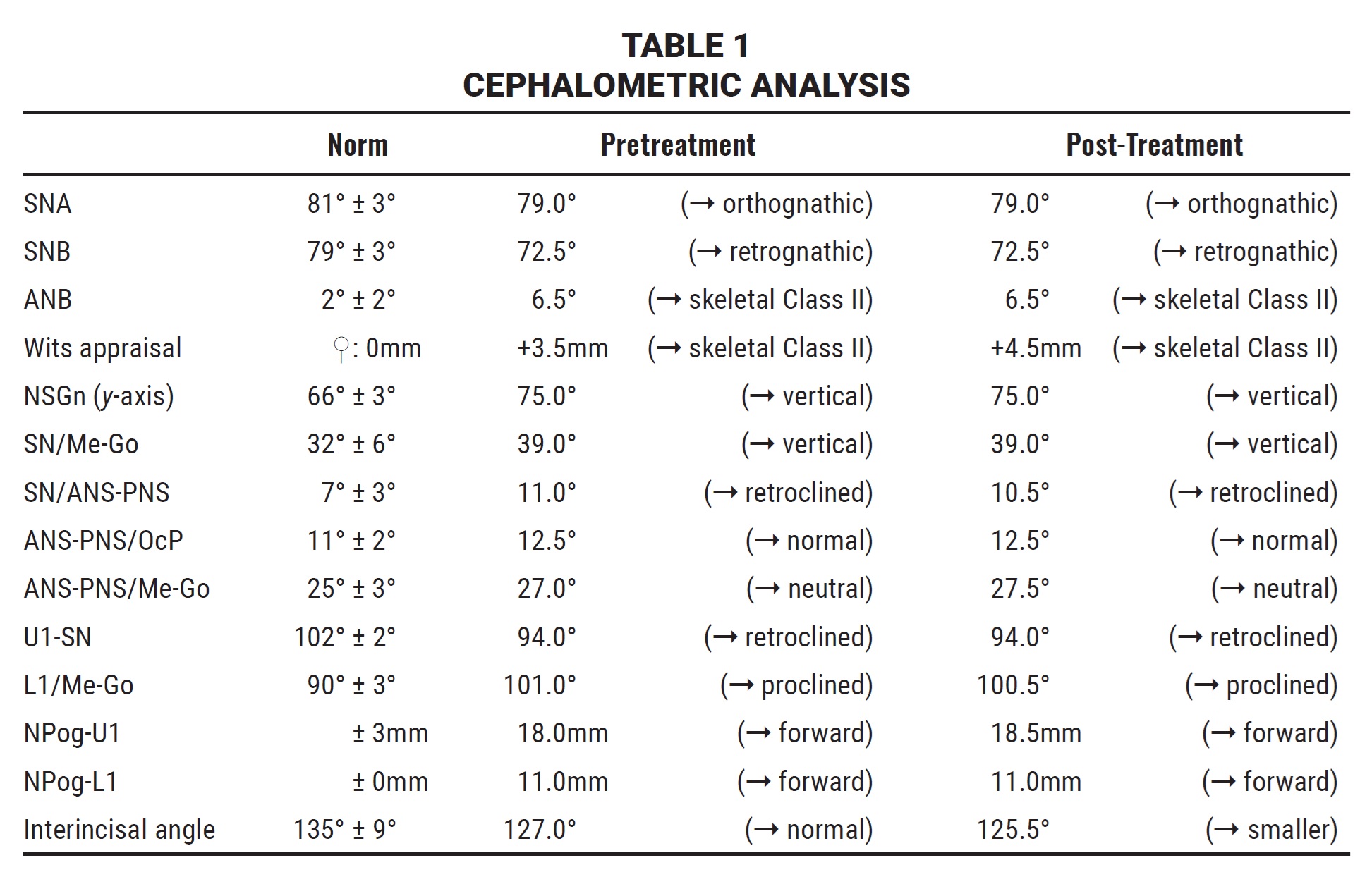
The panoramic radiograph confirmed the patient’s overall dental and periodontal health. Cephalometric analysis (Table 1) indicated a skeletal Class II malocclusion due to a retrognathic mandible (SNB = 72.5°) and a vertical facial type (SN/Me-Go = 39°), along with retroclination of the maxillary base and a steep occlusal plane. The upper incisors were retroclined (U1-SN = 94°), and the lower incisors proclined (L1/Me-Go = 101°), while both were forwardly positioned (NPog-U1 = 18mm, NPog-L1 = 11mm), forming a convex lip profile.
The treatment goal was to restore the lower arch by retroclining the lower left canine and retruding the lower left incisors. Although our plan also addressed the gingival recession, only the orthodontic treatment will be detailed in this article.
The patient requested an invisible appliance, but fixed lingual appliances were ruled out because of the excessive orovestibular discrepancy between the lower left first premolar and canine. This discrepancy and the prominent undercuts, especially of the canine, eliminated conventional thermoformed aligners as a treatment option since removal of the trays from the molds would irreversibly deform them. To overcome these challenges, we planned to 3D-print customized aligners from a material with thermomechanical shape memory, thus enabling aligner insertion and removal while applying the continuous light forces needed to avoid exacerbation of the gingival recession.12
Treatment Progress
Digital planning was performed with Orthosetup*** software using a staging protocol with .6mm of translation and as much as 4° of rotational movement per step (Fig. 2). Nine steps were planned for alignment of the lower anterior teeth; two identical aligners were printed for each step, allowing the trays to be changed weekly and thus limiting any potential force-related consequences of deterioration. Passive attachments were planned for the central buccal aspects of the lower left first premolar and right canine.
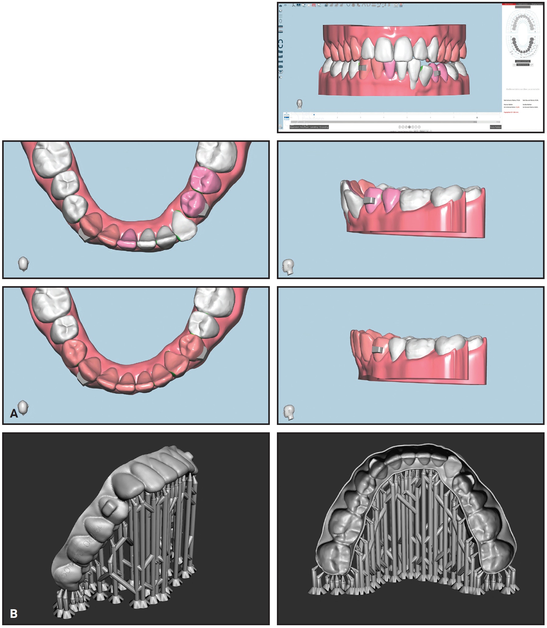
Fig. 2 A. Digital planning in Orthosetup*** software. B. STL aligner shells with supports for 3D printing.
The aligners were printed at a thickness of .6mm from Tera Harz TC-85 photopolymer, using an Asiga Max UV 3D printer and the Tera Harz Cure THC 2 polymerization device. A total of .4mm of interproximal reduction was performed on the mesial and distal sides of the lower left premolars to provide sufficient space for the intended incisor and canine movements.
Treatment progress was monitored every four weeks, with new aligners fabricated in time for each appointment. The patient reported that the aligners were more comfortable than the fixed orthodontic appliance that had been used during her previous treatment.
After the first nine steps had aligned the lower left canine and incisors (Fig. 3), six more aligners were produced for an additional three steps of refinement.
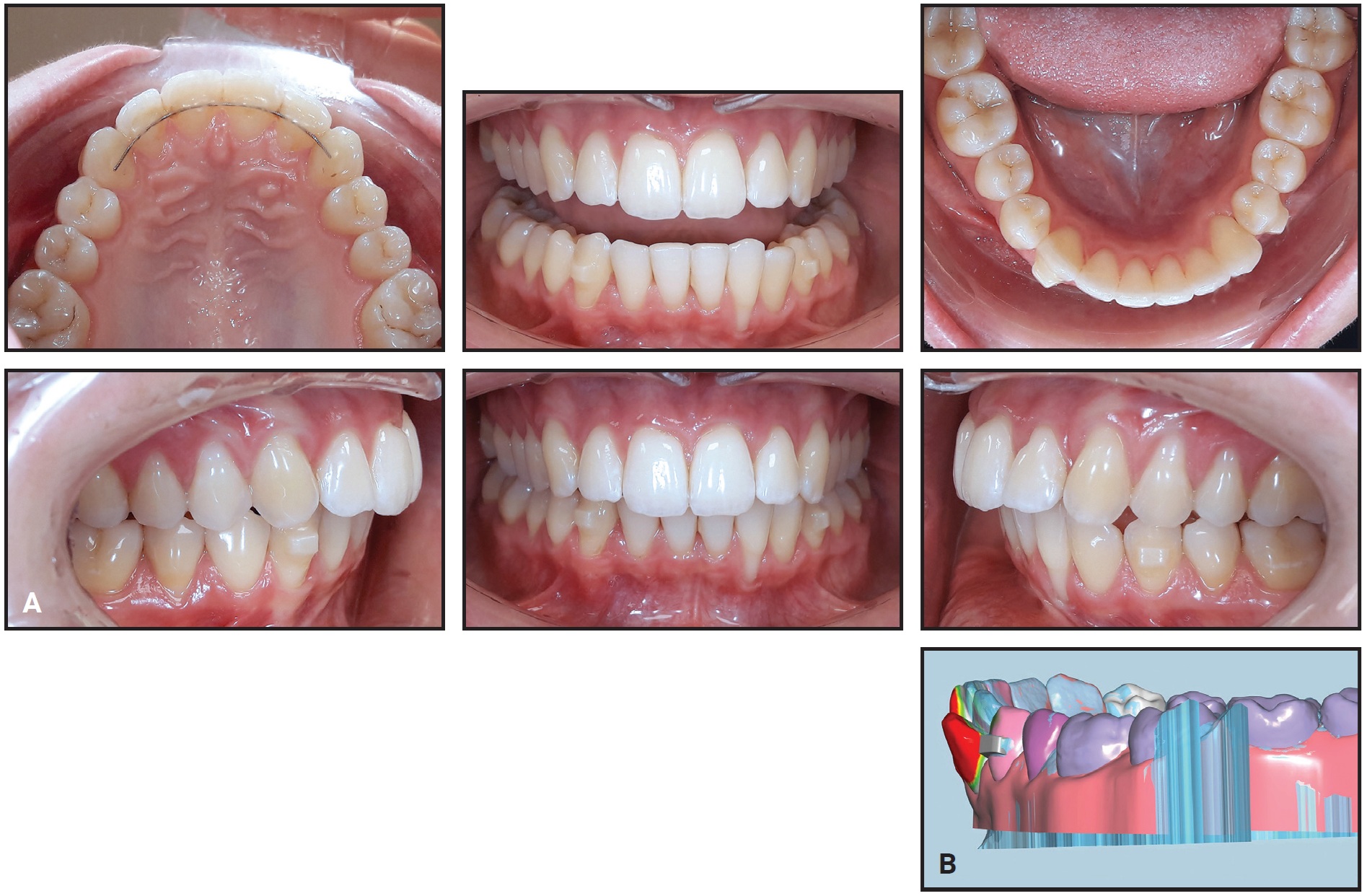
Fig. 3 A. After 18 weeks of treatment with 3D-printed aligners. B. Comparison with pretreatment model (red = areas of greatest change).
Active treatment thus involved 12 steps, with a total of 24 lower aligners worn for 24 weeks (Fig. 4).
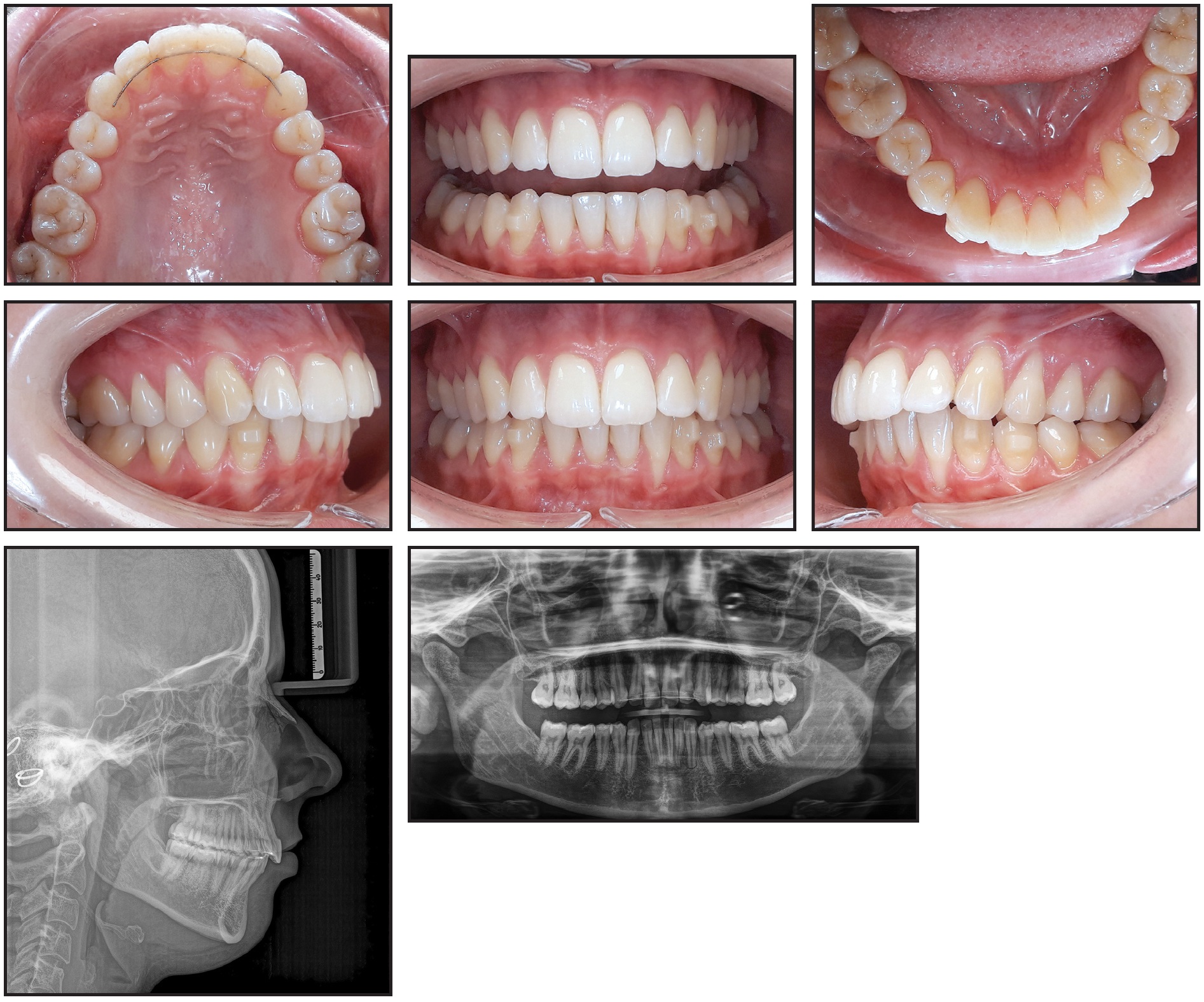
Fig. 4 After 24 weeks, including nine steps of treatment and three steps of refinement.
A removable retainer was delivered for two months of wear, after which a lower 3-3 Nitinol CAD/CAM Memotain† lingual retainer wire was bonded (Fig. 5).
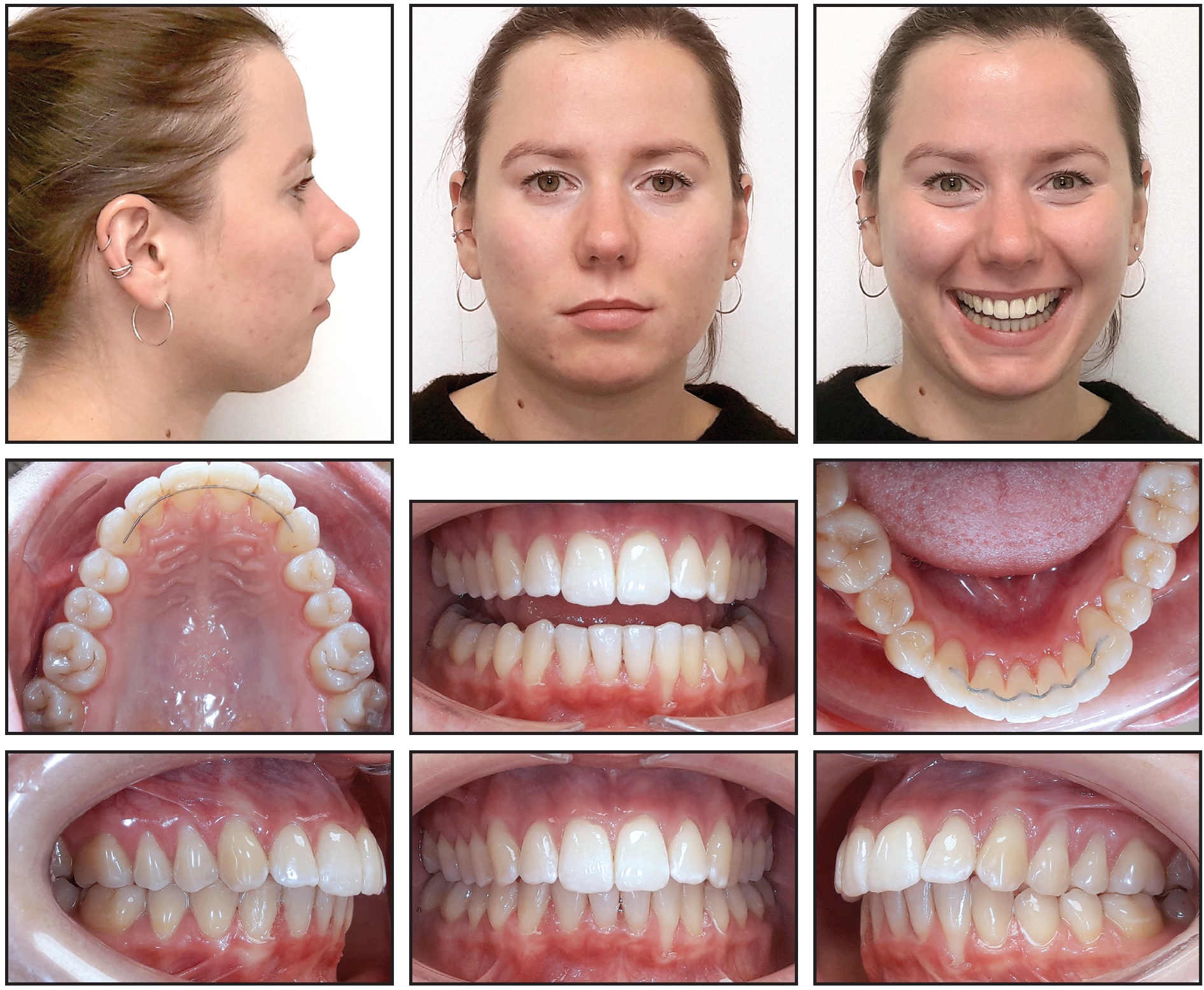
Fig. 5 About eight months after beginning of treatment, with bonded lower 3-3 Memotain† lingual retainer wire in place.
Treatment Results
The protrusion of the lower left incisors and proclination of the lower left canine were corrected, realigning the lower arch and closing the open bite. Controlled lingual tipping of the lower left lateral incisor reduced the depth of the gingival recession from 5mm to 3.5mm and significantly decreased its width. No complications were noted, and the patient was satisfied with the esthetic results. Cephalometric analysis revealed no clinically significant sagittal or vertical changes (Table 1).
Discussion
This article documents the treatment of inadvertent tooth movement caused by the failure of a fixed lingual retainer wire. In contrast to a relapse—a return to the malocclusion corrected by orthodontic treatment—the failed retainer created an even worse malocclusion. The unanticipated torque tension induced by the wire severely compromised the health of the affected teeth. The lower left incisors and canine were affected primarily by the outward rotational force of the retainer wire. For the canine, this force resulted in uncontrolled tipping, which tilted the crown buccally and the root lingually (Fig. 1). When the left end of the wire moved in a buccal direction, it drew the crowns of the lower incisors buccally, while the lingual root torque applied by the retainer kept their apices in about the same location. By tilting the root of the lateral incisor out of the alveolar bone, this movement promoted the development of gingival recession.
When the gingival tissue around a tooth is damaged, it is imperative to apply targeted, consistent forces of low magnitude for traction.12 Unlike standard thermoformed aligners, those printed with TC-85 photopolymer have been shown to adapt fully to the shapes of the incisor crowns,10 enabling targeted application of the necessary light forces.
Our plan also took advantage of the TC-85 material’s shape-memory feature, which enables an aligner to become flexible enough for easy insertion when exposed to hot water but to return to its original shape upon cooling to body temperature. That flexibility also makes the aligners more comfortable to wear.10
Despite the many potential advantages of this new material, the relatively small body of literature regarding its clinical performance may have limited our treatment options. Our aligners were designed to be changed weekly because a study of the ability of 3D-printed TC-85 aligners to maintain their mechanical properties during use tested them for only one week.14 Although the TC-85 resin allows different parts of the aligner to be printed at different thicknesses,9,15 potentially producing force vectors comparable to those achieved with attachments on the teeth,13 we relied on conventional attachments because no evidence-based scientific study had measured those forces in a case similar to ours. Further gaps in the literature include the consequences of errors made during certain steps in the workflow for designing and printing aligners, which can sometimes result in substantial unintended forces and moments.13,16
This case underscores the efficacy of customized, technologically driven approaches in treating complex cases. The unique properties of 3D-printed aligners with shape memory allowed us to save a lower lateral incisor with an impaired periodontium and effectively manage protruded lower incisors and a proclined lower canine without the need for additional skeletal anchorage. As the body of scientific and clinical knowledge about 3D-printed aligners continues to grow, they are likely to become a transformative alternative to standard thermoformed aligners and conventional fixed appliances.
FOOTNOTES
- *Registered trademark of Graphy, Inc., Seoul, Korea; www.itgraphy.com.
- **Registered trademark of Asiga, Alexandria, Australia; www.asiga.com.
- ***Scheu Dental GmbH, Iserlohn, Germany; www.scheu-dental.com.
- †Registered trademark of CA Digital GmbH, Hilden, Germany; www.ca-digit.com.
REFERENCES
- 1. Hartshorne, J. and Wertheimer, M.B.: Emerging insights and new developments in clear aligner therapy: A review of the literature, Am. J. Orthod. Clin. Comp. 2:311-324, 2022.
- 2. Tuncay, O.C.: The Invisalign System, Quintessence Publishing United Kingdom, New Malden, U.K., 2006.
- 3. Alansari, R.A.; Faydhi, D.A.; Ashour, B.S.; Alsaggaf, D.H.; Shuman, M.T.; Ghoneim, S.H.; Linjawi, A.I.; Marghalani, H.Y; and Dause, R.R.: Adult perceptions of different orthodontic appliances, Patient Prefer. Adherence 13:2119-2128, 2019.
- 4. Weir, T.: Clear aligners in orthodontic treatment, Austral. Dent. J. 62:58-62, 2017.
- 5. Sachdev, S.; Tantidhnazet, S.; and Saengfai, N.N.: Accuracy of tooth movement with in-house clear aligners, J. World Fed. Orthod. 10:177-182, 2021.
- 6. Lee, S.Y.; Kim, H.; Kim, H.J.; Chung, C.J.; Choi, Y.J.; Kim, S.J.; and Cha, J.Y.: Thermo-mechanical properties of 3D printed photocurable shape memory resin for clear aligners, Sci. Rep. 12:6246, 2022.
- 7. Koenig, N.; Choi, J.Y.; McCray, J.; Hayes, A.; Schneider, P.; and Kim, K.B.: Comparison of dimensional accuracy between direct-printed and thermoformed aligners, Kor. J. Orthod. 52:249-257, 2022.
- 8. Tartaglia, G.M.; Mapelli, A.; Maspero, C.; Santaniello, T.; Serafin, M.; Farronato, M.; and Caprioglio, A.: Direct 3D printing of clear orthodontic aligners: Current state and future possibilities, Mater. (Basel) 14:1799, 2021.
- 9. Grant, J.; Foley, P.; Bankhead, B.; Miranda, G.; Adel, S.M.; and Kim, K.B.: Forces and moments generated by 3D direct printed clear aligners of varying labial and lingual thicknesses during lingual movement of maxillary central incisor: An in vitro study, Prog. Orthod. 24:23, 2023.
- 10. Park, S.Y.; Choi, S.H.; Yu, H.S.; Kim, S.J.; Kim, H.; Kim, K.B.; and Cha, J.Y.: Comparison of translucency, thickness, and gap width of thermoformed and 3D-printed clear aligners using micro-CT and spectrophotometer, Sci. Rep. 13:10921, 2023.
- 11. Pratsinis, H.; Papageorgiou, S.N.; Panayi, N.; Iliadi, A.; Eliades, T.; and Kletsas, D.: Cytotoxicity and estrogenicity of a novel 3-dimensional printed orthodontic aligner, Am. J. Orthod. 162:e116-e122, 2022.
- 12. Hertan, E.; McCray, J.; Bankhead, B.; and Kim, K.B.: Force profile assessment of direct-printed aligners versus thermoformed aligners and the effects of non-engaged surface patterns, Prog. Orthod. 23:49, 2022.
- 13. McKay, A.; McCray, J.; Bankhead, B.; Lee, M.M.; Miranda, G.; Adel, S.M.; and Kim, K.B.: Forces and moments generated during extrusion of a maxillary central incisor with clear aligners: An in vitro study, BMC Oral Health 23:495, 2023.
- 14. Can, E.; Panayi, N.; Polychronis, G.; Papageorgiou, S.N.; Zinelis, S.; Eliades, G.; and Eliades, T.: In-house 3D-printed aligners: Effect of in vivo ageing on mechanical properties, Eur. J. Orthod. 44:51-55, 2022.
- 15. McCarty, M.C.; Chen, S.J.; English, J.D.; and Kasper, F.: Effect of print orientation and duration of ultraviolet curing on the dimensional accuracy of a 3-dimensionally printed orthodontic clear aligner design, Am. J. Orthod. 158:889-897, 2020.
- 16. Panayi, N.; Cha, J.Y.; and Kim, K.B.: 3D printed aligners: Material science, workflow and clinical applications, Semin. Orthod. 29:25-33, 2023.
-
 DR. SIVAK
 -
 DR. JO
 -
 DR. NANDA
 -
 DR. BECHTOLD
Dr. Sivak is the Director of Product Development, CA Digital GmbH, Hilden, Germany. Dr. Jo is an orthodontist and the Director, Doktor Zahnspange, Hilden, Germany. Dr. Nanda is an Associate Editor of the Journal of Clinical Orthodontics; Professor Emeritus, University of Connecticut, Farmington, CT; and an Adjunct Professor, ADA Forsyth Institute, Cambridge, MA. Dr. Bechtold is an orthodontist and an Associate Professor, Department of Prosthodontics, Geriatric Dentistry and Craniomandibular Disorders/Section of CMD, Campus Benjamin Franklin, Center for Dental and Craniofacial Sciences (CC3), Charité-Universitätsmedizin Berlin, Corporate Member of Freie Universität Berlin, Humboldt-Universität zu Berlin, and Berlin Institute of Health, Assmannshauser Strasse 4-6, 14197 Berlin, Germany; e-mail: till.bechtold@charite.de.