Favorite Saved

A Reinforced Clear Retainer for the Mixed Dentition

Maxillary retainers are sometimes used in the mixed dentition after treatment of anterior crossbite or unerupted maxillary incisors.1 Since the deciduous molars are usually replaced by permanent teeth during this period, a Hawley-type retainer must be adjusted to allow for eruption of the premolars. A properly designed thermoplastic retainer is much more economical than an acrylic retainer and can usually be fabricated in the office and placed on the same day, without the need for a commercial laboratory service.2

In 2003, Theroux described a vacuum-formed Phase I retainer made from double layers of Biocryl* sandwiched around a layer of light-cured acrylic.3 The first Biocryl layer was adapted to the cast on the vacuum-forming machine and cut out in the shape of the palate. The light-cured acrylic was then applied in a horseshoe shape, and the final Biocryl layer was thermoformed over the previous layers. A major disadvantage of this design is the time involved in fabrication; in addition, the author later mentioned that fractures could occur around the first molars due to the single thickness of thermoformed material in those regions.4

Similar articles from the archive:

At our university, we have developed an acrylic-reinforced clear retainer for which the thermoplastic material is formed directly over chemically cured acrylic, saving steps and fabrication time.

Fabrication

1. After removal of the Phase I fixed appliances, take an alginate impression of the maxillary arch and pour it in stone.

2. Apply a separating medium** to the cast in the palate and other areas where acrylic will be added, and allow it to dry for five to 10 minutes.

3. Apply acrylic powder and liquid to the same areas of the cast, using the "salt and pepper" technique. Do not allow the acrylic to cure, or it will not adhere to the thermoplastic material. Apply the final coat while the thermoplastic sheet (1mm Biocryl) is still preheating.

4. Place the Biocryl sheet over the stone cast in a vacuum-forming machine, and suck down the clear retainer. Allow the acrylic to cure for 15 minutes.

5. Trim away the thermoplastic material from the deciduous-molar areas to allow their exfoliation during the retention period (Fig. 1). To reduce the risk of fracture in the first-molar areas, the thermoplastic material covering the second deciduous molars can be left in place and removed as necessary at later appointments. Alternatively, a thicker, 1.5mm Biocryl sheet can be used.

Clinical Management

An 8-year-old male patient with rotated maxillary incisors and an anterior crossbite was treated with a 2 x 4 fixed appliance (Fig. 2A). The maxillary left first premolar was already erupting through the gingiva at the end of Phase I (Fig. 2B). To maintain the positions of the maxillary incisors, a clear reinforced retainer was fabricated as described above, with the thermoplastic material removed from the canine and deciduous-molar regions (Fig. 2C). The patient was seen every four to six months, and the edges of the thermoplastic material were trimmed as needed, using an acrylic bur in a slow-speed handpiece, to allow eruption of the premolars.

Letting the patient choose the acrylic color seems to promote compliance. The retainer is seated in the mouth with finger pressure, and the patient is shown how to remove the appliance by pulling down on the incisal material. The patient is usually instructed to wear the retainer only while sleeping, to store it in a retainer case when it is not in the mouth, and to brush it daily with toothpaste.

FOOTNOTES

Fig. 1 Reinforced clear retainer for mixed-dentition patient. Thermoplastic material is cut away to allow exfoliation of permanent canines and first premolars, while second deciduous molars remain covered to help prevent acrylic fracture in first-molar regions.

Fig. 2 A. 8-year-old male patient before early treatment of anterior crossbite. B. Maxillary left first premolar erupting at end of Phase I. C. Reinforced clear retainer, with canine and deciduous-molar regions trimmed away to allow eruption of permanent dentition.

REFERENCES

  • 1.   Dugoni, S.A.; Lee, J.S.; Varela, J.; and Dugoni, A.A.: Early mixed dentition treatment: Post-retention evaluation of stability and relapse, Angle Orthod. 65:311-320, 1995.
  • 2.   Sheridan, J.J.; LeDoux, W.; and McMinn, R.: Essix retainer: Fabrication and supervision for permanent retention, J. Clin. Orthod. 27:37-45, 1993.
  • 3.   Theroux, K.L.: A new vacuum-formed Phase I retainer, J. Clin. Orthod. 37:384-387, 2003.
  • 4.   Theroux, K.L.: A modified Theroux Phase I retainer design, J. Clin. Orthod. 45:666-668, 2011.
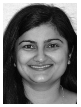
  • DEEPA
    DR. VYAS
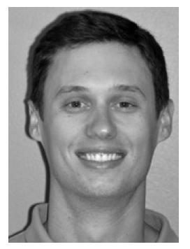
  • NICHOLAS
    DR. MADDUX
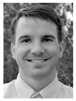
  • TODD
    DR. BOVENIZER
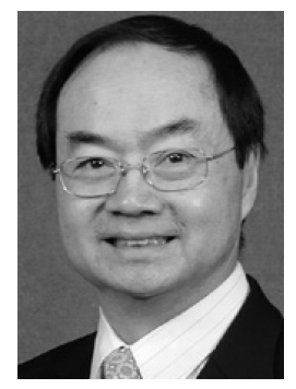
  • PETER
    DR. NGAN

Drs. Vyas and Maddux are orthodontic residents and Dr. Ngan is Professor and Chair, Department of Orthodontics, School of Dentistry, West Virginia University, Robert C. Byrd Health Sciences Center, Morgantown, WV 26506. Dr. Bovenizer is in the private practice of orthodontics in Cary, NC. E-mail Dr. Ngan at pngan@hsc.wvu.edu.

Fig. 1 Reinforced clear retainer for mixeddentition patient. Thermoplastic material is cut away to allow exfoliation of permanent canines and first premolars, while second deciduous molars remain covered to help prevent acrylic fracture in first-molar regions.
Fig. 2 A. 8-year-old male patient before early treatment of anterior crossbite. B. Maxillary left first premolar erupting at end of Phase I. C. Reinforced clear retainer, with canine and deciduous- molar regions trimmed away to allow eruption of permanent dentition.

FOOTNOTES

REFERENCES 2

DR. DEEPA VYAS DMD

DR. DEEPA  VYAS DMD

DR. NICHOLAS MADDUX DDS

DR. NICHOLAS  MADDUX DDS

DR. TODD BOVENIZER DDS, MS

DR. TODD  BOVENIZER DDS, MS

DR. PETER NGAN DMD

DR. PETER  NGAN DMD

My Account

This is currently not available. Please check back later.

Please contact heather@jco-online.com for any changes to your account.