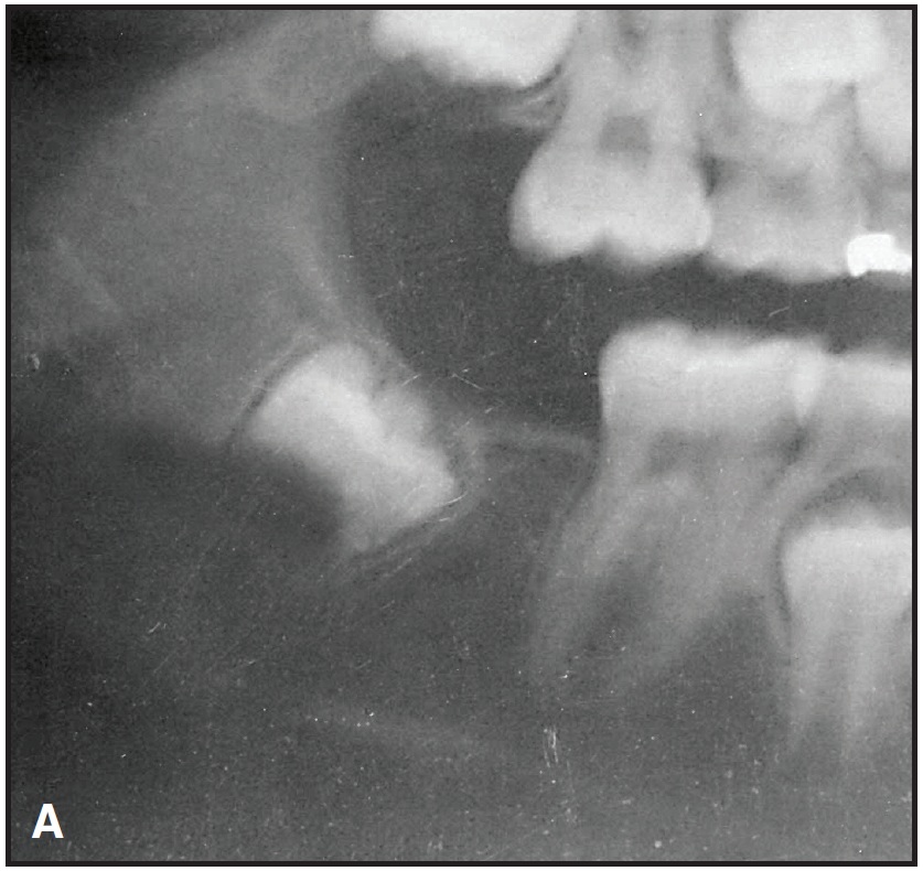
An 11-year-old female presented for orthodontic treatment. The records were unremarkable save for one thing: the panoramic x-ray showed an extraordinarily wide span from the distal of the mandibular right first molar to the mesial of the mandibular right second molar (A). It wasn't until the mandibular left second molar had almost fully erupted about a year later, with no sign of the mandibular right second molar, that a red flag was raised.
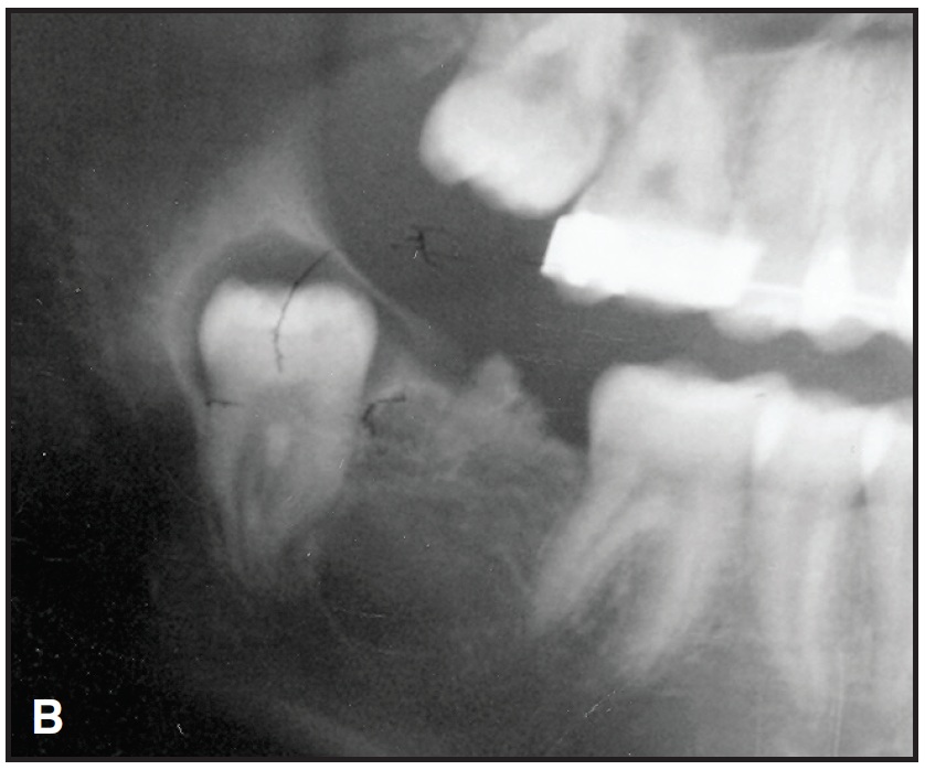
A progress panorex then displayed what appeared to be an odontoma (B), which was confirmed by biopsy. The x-ray also showed that the mandibular right second molar had fully formed, but had drifted so far distally that it had begun erupting superiorly into the ascending ramus. The crown of the tooth was surrounded by what appeared to be a dentigerous cyst.
Similar articles from the archive:
- Modified Herbst Appliance for the Mixed Dentition November 1985
The surgeon determined that the best course of action was to remove the wayward second molar, the surrounding cyst, and the odontoma. All went well, and healing was rapid and uneventful.
My lesson learned: take progress films on all eruption aberrancies observed in the initial records, no matter how insignificant they may seem.
-
 DR. GOODMAN
Dr. Goodman is in the private practice of orthodontics, 30 W. Rahn Road, Dayton, OH 45429. E-mail him at: pmarshallg@live.com.