Correction of a Canted Lower Incisal Plane
A cant of the mandibular incisal plane can be difficult to recognize in a patient with a unilateral skeletal crossbite due to a lateral CO-CR shift, especially if the patient has an impinging deep bite. A differential diagnosis between an incisal cant and an occlusal cant is of primary importance, because while the former can be corrected with well-controlled, determinate force systems,1-3 the latter usually requires controlling the eruption of the buccal segments in growing individuals or, in adults, can often be resolved only through ortho-gnathic surgery.4,5
This article demonstrates an effective method by which an incisal cant can be leveled to achieve an ideal occlusion with a symmetrical overbite.
Case Report
A 12-year-old male in the permanent dentition presented with the chief complaint of an anterior crossbite. He had a Class II, division 1 subdivision right malocclusion with a narrow maxilla (Fig. 1). The maxillary midline coincided with the facial midline, but the mandibular midline had shifted 3mm to the right, resulting in a unilateral crossbite. The patient also showed a slight cant of the lower incisor segment.
Similar articles from the archive:
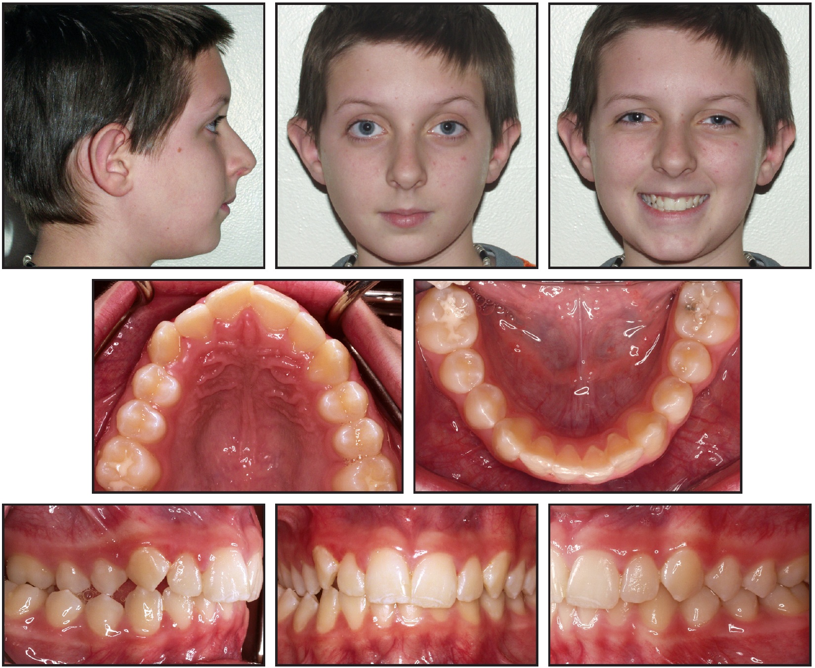
Fig. 1 12-year-old male patient with maxillary constriction and unilateral crossbite due to mandibular shift to right. Mandibular incisal cant is camouflaged by crossbite.
Comprehensive orthodontic treatment was begun with rapid palatal expansion of the maxillary arch. Full fixed appliances were then bonded for leveling and alignment, with a continuous .014" nickel titanium wire placed in the lower arch. At that point, a significant mandibular cant became obvious (Fig. 2). One month later, an .016" x .022" stainless steel archwire was sectioned between the canines and incisors on both sides, and an .017" x .025" CNA Beta Titanium* cantilever wire was bent down into the mucobuccal fold on the left side, from the first molar auxiliary tube to the main archwire between the central and lateral incisors (Fig. 3).
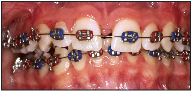
Fig. 2 Lower incisal cant evident after maxilla was expanded and initial leveling archwires were placed.
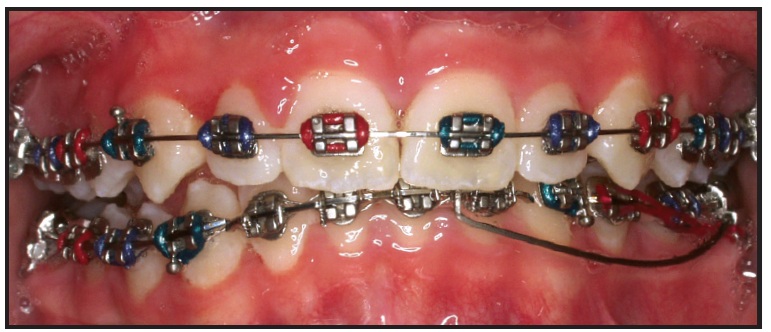
Fig. 3 Lower .016" ✕ .022" stainless steel archwire sectioned distal to lateral incisors; .017" ✕ .025" CNA Beta Titanium cantilever wire extended from lower left first molar to anterior segment.
This point of attachment, off-center from the lower midline, was selected to obtain not only an intrusive force, but a moment around the center of resistance of the anterior segment (Fig. 4). To prevent any spaces from opening, the segments from the lower right first molar to the left lateral incisor and from the lower left first molar to the left canine were ligated together.
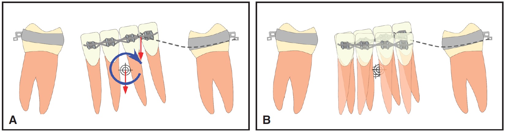
Fig. 4 A. Applied force system. B. Effect of cantilever producing off-center intrusive force on canted lower incisal plane.
After 10 weeks, with no reactivation of the cantilever, a significant amount of intrusion and rotation of the anterior segment had been achieved, but the incisal cant was not fully corrected. The cantilever was then moved to the distal end of the anterior segment, thus increasing the magnitude of the moment in relation to the center of resistance of the anterior teeth (Fig. 5).
Once the lower anterior segment had been corrected, a vertical discrepancy between the incisors and the left canine became evident. A similar cantilever with an intrusive force was then attached to the lower left canine, and an .016" x .022" stainless steel archwire was inserted from the lower right first molar to the lower left lateral incisor to maintain the correction of the incisal cant (Fig. 6).
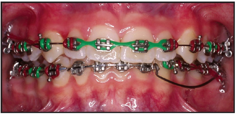
Fig. 5 Cantilever moved to distal end of lower anterior segment to produce greater anterior moment.
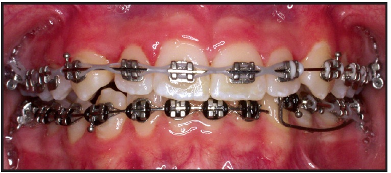
Fig. 6 Cantilever with intrusive force attached to lower left canine to correct vertical discrepancy between lower left canine and incisors.
When the lower arch was almost completely leveled, a continuous .017" x .025" CNA archwire was placed in the lower arch, and a vertical seating elastic was used on the right side to extrude the lower canine and premolar and finish the correction of the incisal cant (Fig. 7). An .017" x .025" stainless steel wire was placed in the upper arch to minimize any extrusion of the maxillary teeth. Detailing was completed with esthetic wire bends (Fig. 8).
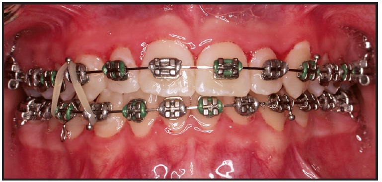
Fig. 7 Vertical elastic worn on right side to complete correction of lower incisal cant.
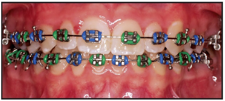
Fig. 8 Patient after full correction of incisal cant, showing coincident midlines; finishing bends placed in .017" ✕ .025" CNA archwire.
The total treatment time was 23 months (Fig. 9).
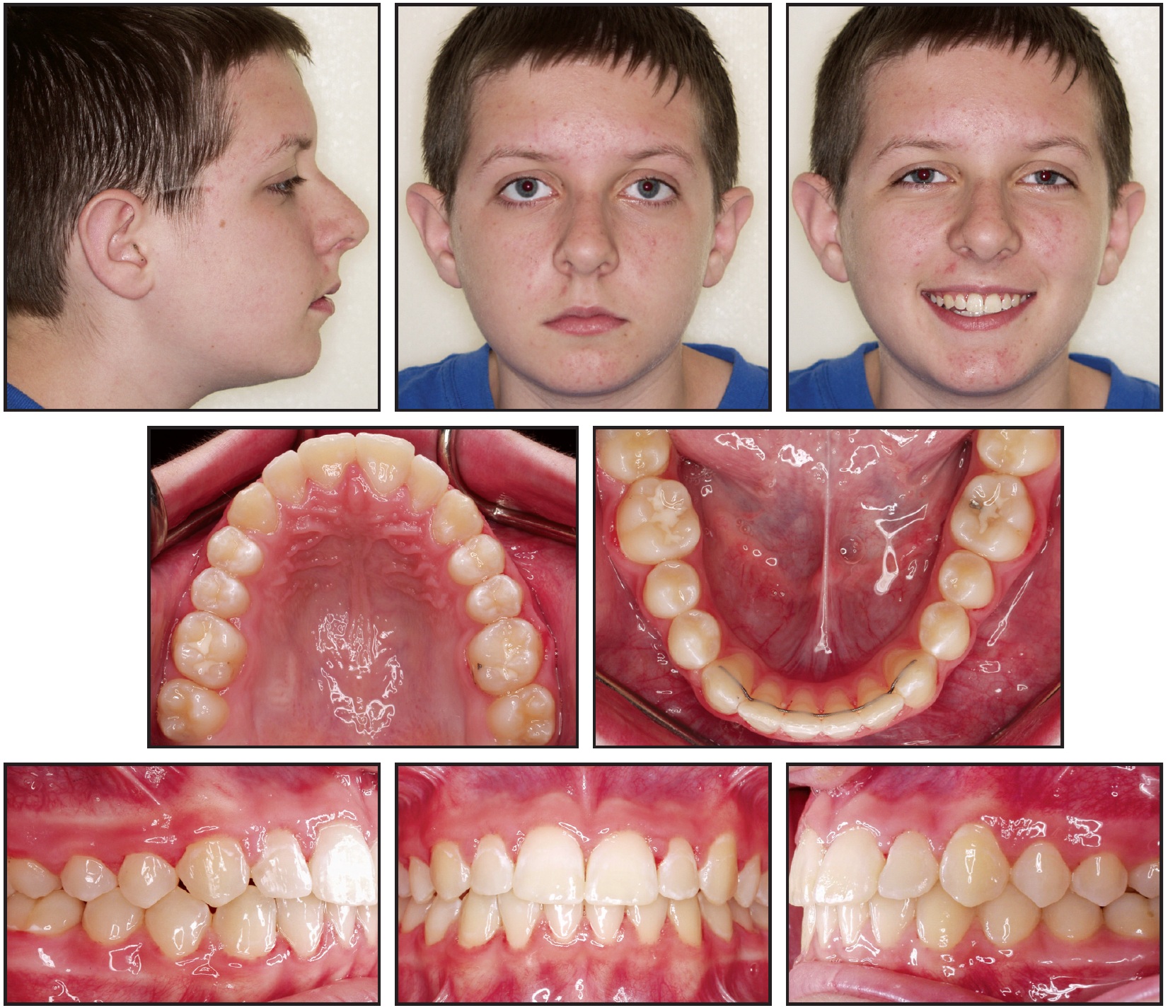
Fig. 9 Patient after 23 months of treatment.
Discussion
Figure 10 shows another patient with a similar lower incisor cant. The same mechanics were applied, using a cantilever to intrude the lower left anterior segment and subsequently intrude the lower left canine.
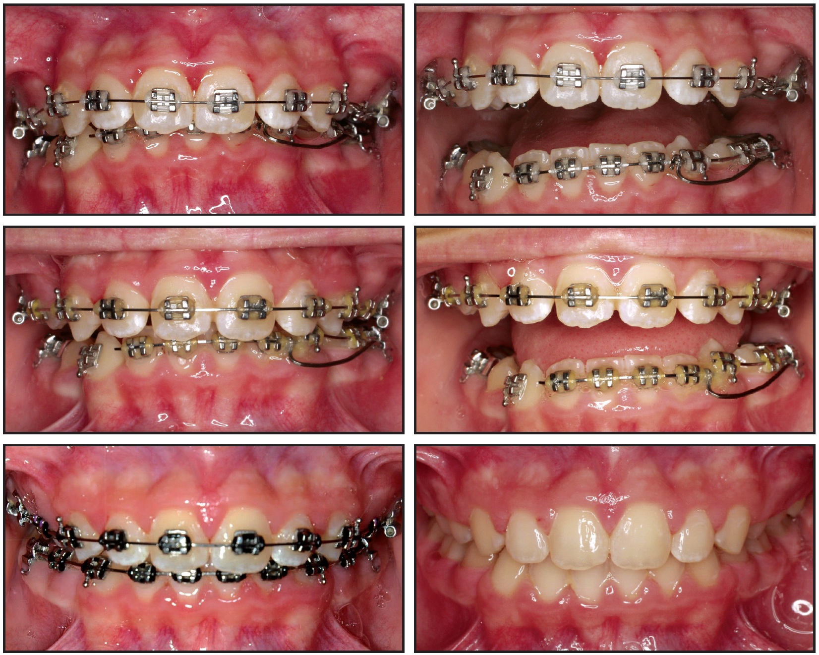
Fig. 10 Similar patient with canted lower incisal plane treated with same mechanics. Cantilever was used to correct inclination of lower incisal plane and then to intrude lower left canine (see Fig. 6).
Segmented arches and cantilevers can effectively correct problems, such as a canted incisal plane, that would be difficult to resolve by any other means. By understanding and controlling side effects, the clinician can achieve an ideal result in a relatively short time.
FOOTNOTES
- *Ortho Organizers, Inc., 1822 Aston Ave., Carlsbad, CA 92008; www.orthoorganizers.com.
REFERENCES
- 1. Legan, H. and Conley, R.: Biomechanical factors in surgical orthodontics, in Biomechanics and Esthetic Strategies in Clinical Orthodontics, ed. R. Nanda, Elsevier, St. Louis, 2005, pp. 310-329.
- 2. Nanda, R. and Margolis, M.J.: Treatment strategies for midline discrepancies, Semin. Orthod. 2:84-89, 1996.
- 3. Nanda, R. and Uribe, F.: Individualized orthodontic treatment planning, in Biomechanics and Esthetic Strategies in Clinical Orthodontics, ed. R. Nanda, Elsevier, St. Louis, 2005, pp. 74-93.
- 4. Burstone, C. and Marcotte, M.: The treatment occlusal plane, in Problem Solving in Orthodontics, Quintessence, Chicago, 2000, pp. 31-50.
- 5. Proffit, W. and Turvey, T.: Dentofacial asymmetry, in Contemporary Treatment of Dentofacial Deformity, ed. W. Proffit, R. White, and D. Sarver, Elsevier, St. Louis, 2003, pp. 574-644.
-
 DR. DELUKE
 -
 DR. URIBE
 -
 DR. NANDA
Dr. DeLuke is in the private practice of orthodontics in Schenectady, NY. Dr. Uribe is Assistant Professor and Program Director, Division of Orthodontics, and Dr. Nanda is Professor and Head, Department of Craniofacial Sciences, Alumni Endowed Chair, School of Dental Medicine, University of Connecticut, Farmington, CT 06032. Dr. Nanda is also an Associate Editor of the Journal of Clinical Orthodontics. E-mail him at nanda@nso.uchc.edu.