CLINICAL AID
Early Lightwire Treatment of Bilateral Impacted Mandibular Cuspids
This article shows a patient with ectopic cuspids treated early with the lightwire technique and uncontrolled forces and finished with bidimensional edgewise appliances.
Diagnosis and Treatment Plan
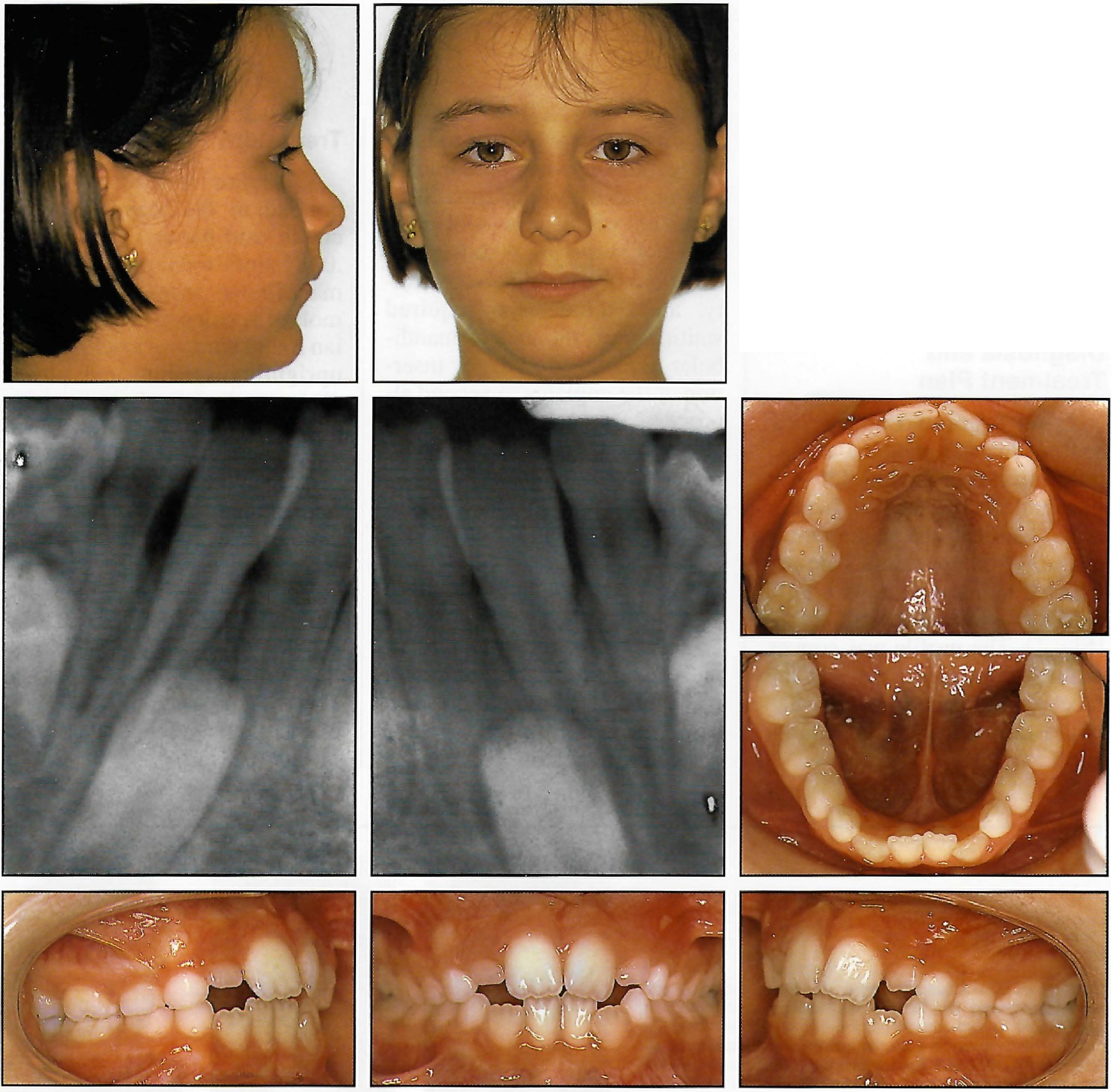
An 8-year-old female presented with the mandibular cuspid crowns positioned between the central and lateral incisor roots. Clinical and radiographic examination revealed a dental Class II malocclusion with mild anterior crowding in both arches (Fig. 1). The profile was convex with a prominent chin, but the face was balanced overall. She had a noticeable tongue-thrust habit.
The patient refused to have the mandibular cuspids extracted and replaced by implants. Such treatment would have involved at least two surgeries, with the possible loss of lateral incisor vitality, and would have required waiting until the end of mandibular alveolar growth for insertion of the implants. A second alternative was to extract the lateral incisors and replace them with the impacted cuspids, but this option was also rejected. Aligning the mandibular lateral incisors and cuspids in transposed positions would have required significant crown recontouring.
Similar articles from the archive:

Fig. 1 8-year-old female patient with mandibular cuspid crowns positioned between roots of central and lateral incisors.
After considering the alternatives, orthodontic treatment was planned to bring the mandibular cuspids into their proper positions in the arch.
Treatment Progress
Lightwire brackets were bonded to the mandibular incisors, and bands with .022" ×.028" slots were placed on the mandibular second deciduous molars. A stopped .018" Australian round wire was inserted, with uprighting springs and power chain applied to the lateral incisors to avoid distal crown movements (Fig. 2).
One month later, the mandibular cuspids were surgically exposed, and metal buttons were bonded to their labial surfaces. An .018" Australian wire with "zig-zag" loops distal to the cuspids was placed, and an elastic chain was applied from the first molars to the cuspids to exert a force with buccal and distal vectors (Fig. 3).
The cuspid crowns were brought into occlusion in another four months. The buttons were then replaced with lightwire brackets, and uprighting springs were inserted to move the roots distally (Fig. 4). The cuspid crowns were stabilized by stainless steel ligatures tied to the permanent molars. Phase I early treatment was completed in 16 months.
Once the mandibular cuspids reached their proper positions and root inclinations, periodontal surgery was performed on their buccal aspects to provide a better quality and quantity of attached gingiva. In the permanent dentition, seven months of Phase II treatment was performed with bidimensional edgewise appliances to correct the Class II malocclusion and produce an ideal intercuspation (Fig. 5).

Fig. 2 Stopped .018" Australian round wire with uprighting springs and power chain applied to mandibular lateral incisors.

Fig. 3 .018" Australian wire with “zig-zag” loops distal to cuspids and elastic traction with buccal and distal vectors.

Fig. 4 Lightwire brackets replacing buttons on cuspids, with uprighting springs used to move roots distally and crowns stabilized by stainless steel ligatures tied to molars.

Fig. 5 Detailing with bidimensional edgewise appliances in permanent dentition.
Treatment Results
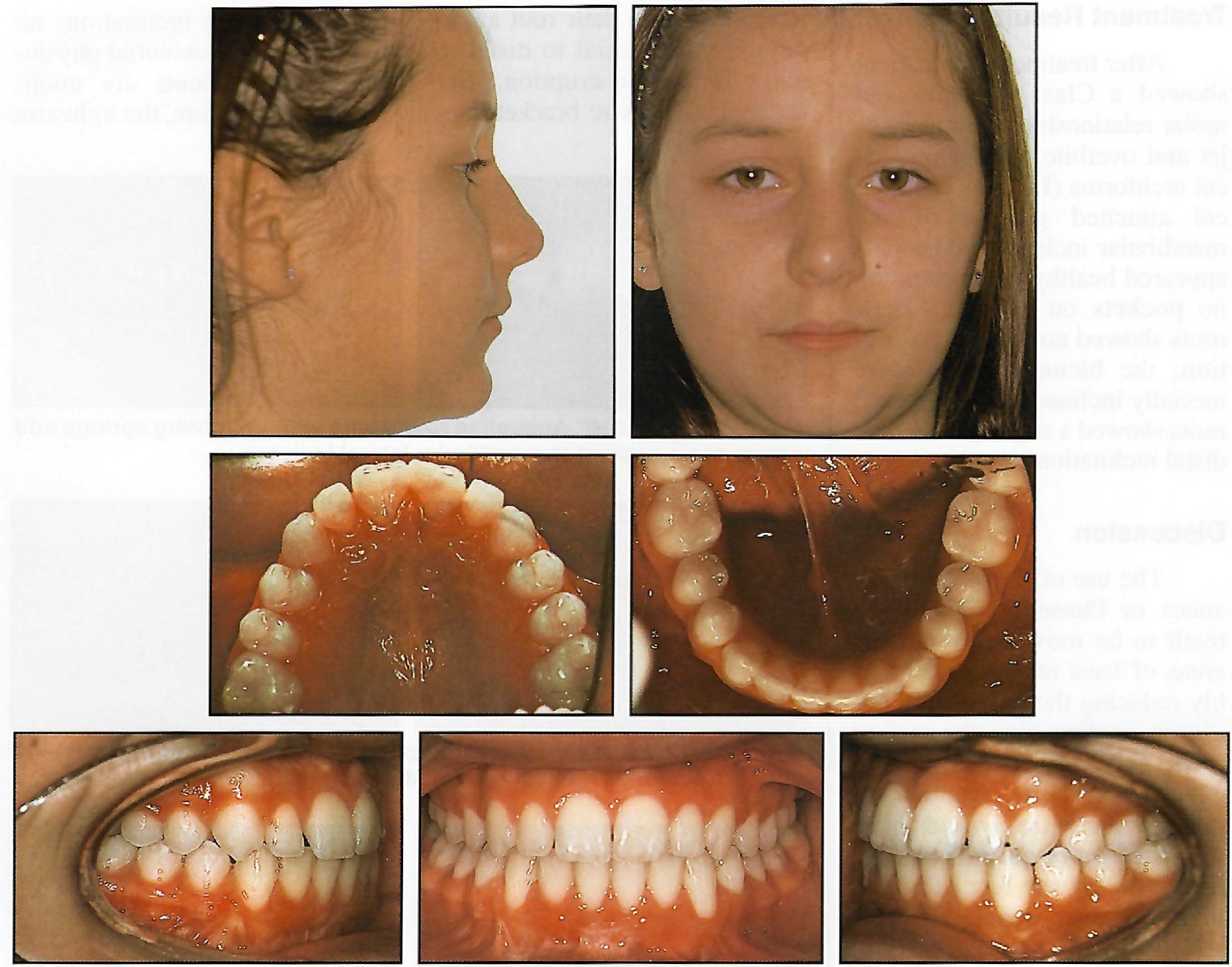
After treatment, the patient showed a Class I cuspid and molar relationship, normal overjet and overbite, and symmetrical archforms (Fig. 6). The buccal attached gingiva of the mandibular incisors and cuspids appeared healthy, demonstrating no pockets on probing. Their roots showed no signs of resorption; the bicuspid roots were mesially inclined, and the cuspid roots showed a slightly excessive distal inclination.

Fig. 6 Patient after Phase II treatment, with mesially inclined mandibular bicuspid roots and distally inclined cuspid roots.
Discussion
The use of lightwire appliances in Phase I allowed the teeth to be moved through the areas of least resistance, probably reducing the risk of impinging on or damaging the incisor roots and cortical bone. With edgewise brackets, even an undersize archwire would have produced more mesiodistal tipping, forcing the teeth through high-resistance areas.
Edgewise bracket prescriptions were developed from permanent dentition cases1-3; in the mixed dentition, the physiological tooth inclination, in-out, and torque are different. In the second transition period, the lower incisors change their root angulations from mesial to distal to allow for cuspid eruption.4 Because the lightwire bracket does not impose any inclinations on the teeth, these essential physiological movements are unobstructed. Therefore, the lightwire technique may be ideal in the mixed dentition, before the permanent teeth have to be taken under control with three-dimensional frictional forces.5-7
ACKNOWLEDGMENT: Surgical treatments were performed by Dr. Carlo Clauser.
REFERENCES
- 1. Andrews, L.F.: The six keys to normal occlusion, Am. J. Orthod. 62:296-307, 1972.
- 2. Andrews, L.F.: The Straight-Wire Appliance, origin, controversy, commentary, J. Clin. Orthod. 10:99-114, 1976.
- 3. Andrews, L.F.: The Straight-Wire Appliance, explained and compared, J. Clin. Orthod. 10:174-195, 1976.
- 4. Van der Linden, F.P.G.M.: Development of the Dentition, Quintessence Publishing Co., Inc., Chicago, 1983.
- 5. Rosa, M.; Cozzani, M.; and Cozzani, G.: Sequential slicing of lower deciduous teeth to resolve incisor crowding, J. Clin. Orthod. 27:596-599, 1994.
- 6. Cozzani, P.; Rosa, M.; and Cozzani, M.: Spontaneous permanent molar expansion in crossbite and non-crossbite patients, abstract of lecture, 75th European Orthodontic Society Congress, Eur. J. Orthod. 21:434, 1999.
- 7. Cozzani, M.; Rosa, M.; Cozzani, P.; and Siciliani G.: Deciduous dentition-anchored rapid maxillary expansion in crossbite and non-crossbite mixed dentition patients: Reaction of the permanent first molar, Prog. Orthod. 4:15-22, 2003.


