The Crown Bracket Bonding System
Crown tip and the vertical position of the teeth in relation to the lips are important elements of the esthetic results achieved by orthodontic treatment.1,2 Crown angulation, in turn, depends on proper placement of the brackets on the crowns from the beginning of treatment.
Tip is the angle between the occlusal plane and the crown long axis. When tip was first built into the bracket body by manufacturers, the slot was milled at an angle, making it difficult to place the bracket accurately in relationship to the occlusal plane and the crown long axis. With the rhomboidal bracket configuration*, developed in 1983, the mesial and distal bracket wings could be used to align the bracket parallel to the long axis. In 1991, the trapezoidal Elite** bracket related the mesial and distal bracket contours to the tip of the root.
Now a new bracket placement method, the Crown System (patent pending), has been developed for use with both Time*** and Crown Line† brackets. This system is based on the principle of congruent surfaces--in other words, surfaces that have the same geometric form, but different sizes. For example, if you try to center a smaller coin on a larger one (Fig. 1), it is easy to recognize with the naked eye when the coins are off-center. Even if a segment of the larger coin is missing, the principle of congruent surfaces still works. The same concept can be applied to the relationship between a bracket and the crown of a tooth.
Similar articles from the archive:
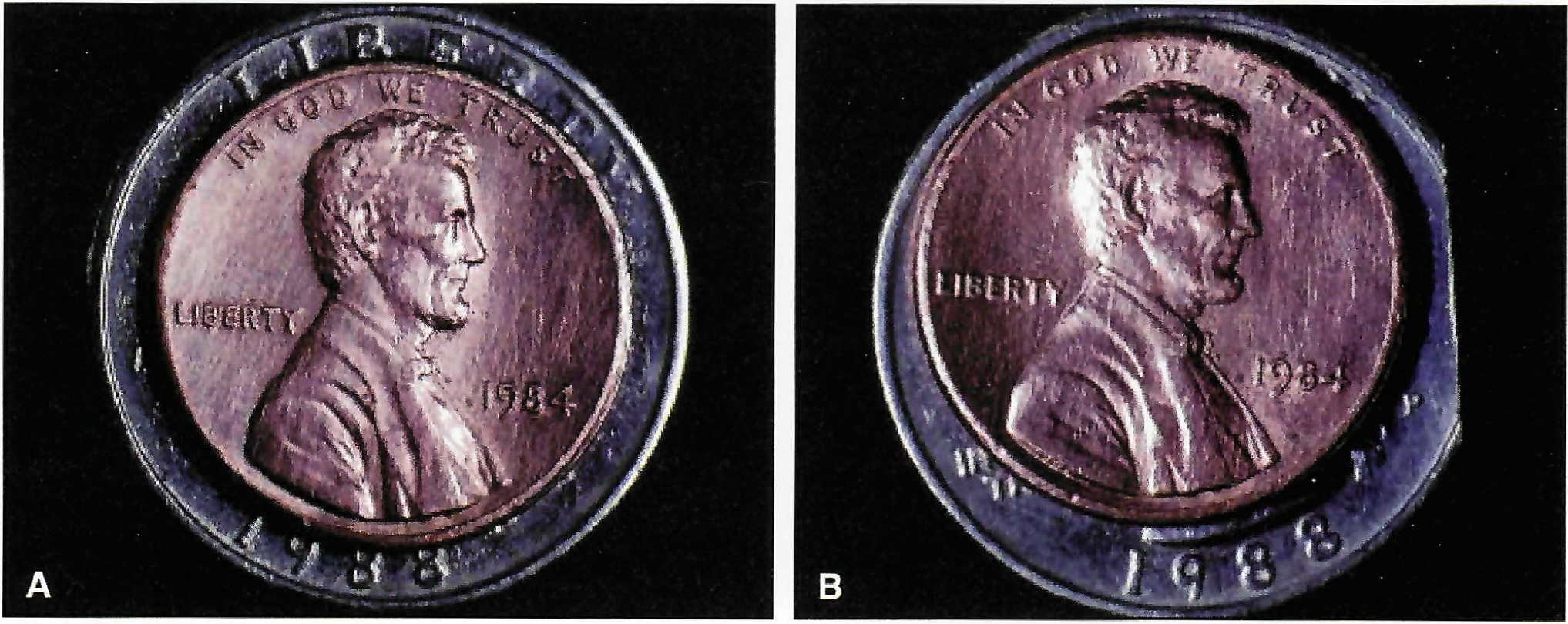
Fig. 1 A. Principle of congruent surfaces used to align coins of different sizes. B. Even if part of larger coin is missing, it is still obvious when smaller coin is off-center.
Analysis of Crown Forms
To determine the correct mean crown forms, intact teeth were photographed from the buccal and the lingual and then enlarged 4.5 times. The mesial and distal crown secants were drawn for each tooth, starting at the intersection between the incisal edge and the distal or mesial crown crest, and ending at the cemento-enamel junction (Fig. 2). Since the cemento-enamel junction cannot be seen in a patient with healthy gingivae or established with a probe, its location was estimated. The crown long axis and the occlusal plane were also constructed for each tooth.
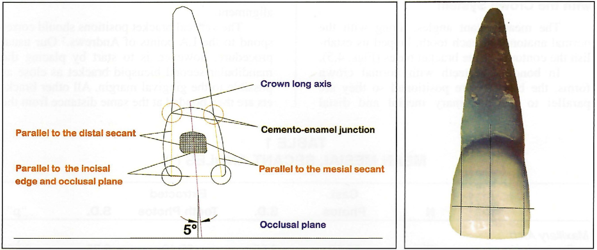
Fig. 2 Mesial and distal secants, crown long axis, and occlusal plane drawn on enlarged photograph of maxillary central incisor.
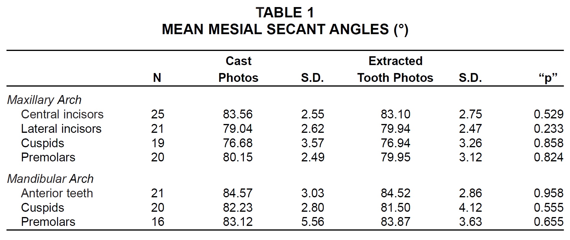
The start and endpoints of the secants were digitized, and the angles formed by these lines with the occlusal plane were calculated with a graphic software program, AutoCAD 12. The means, medians, and standard deviations of the angles were calculated using SPSS for Windows.
To test the accuracy and reproducibility of the estimated cemento-enamel junction locations of the secant endpoints, a second group of teeth was evaluated. In this sample, the secants, crown long axes, and occlusal planes were drawn on photographs of plaster casts (Fig. 3).
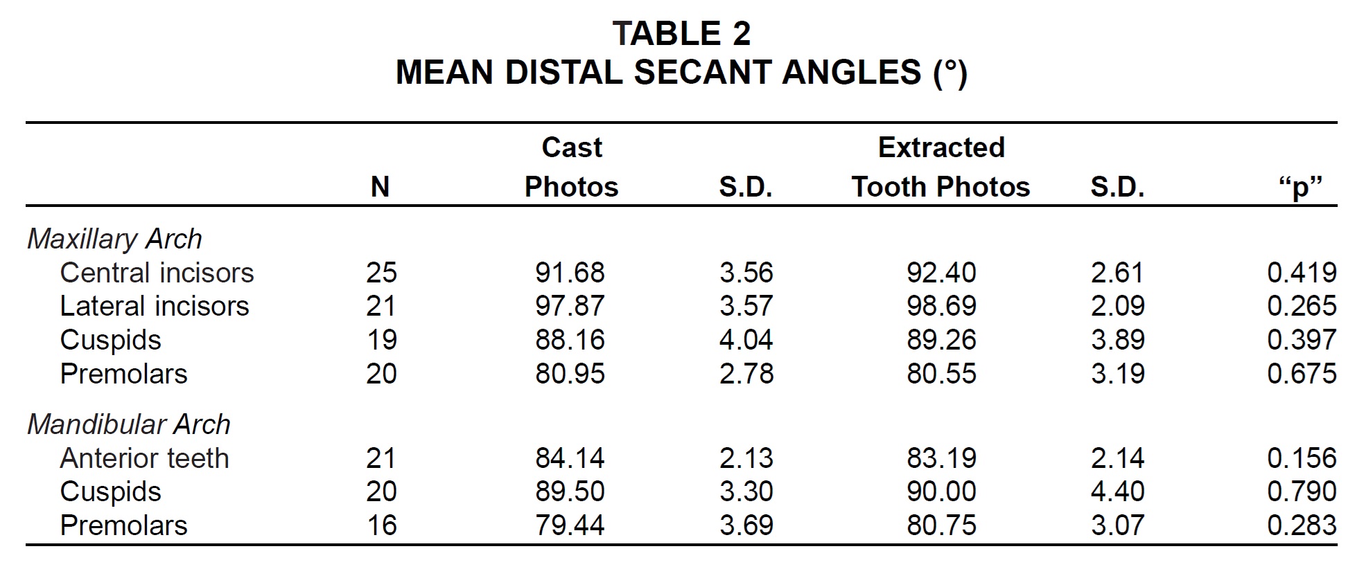
Analysis of variance and an independent-samples t-test were carried out to evaluate the differences among the means, with statistical significance established at the .05 level. No significant difference could be found between the two measurement methods (Tables 1 and 2).
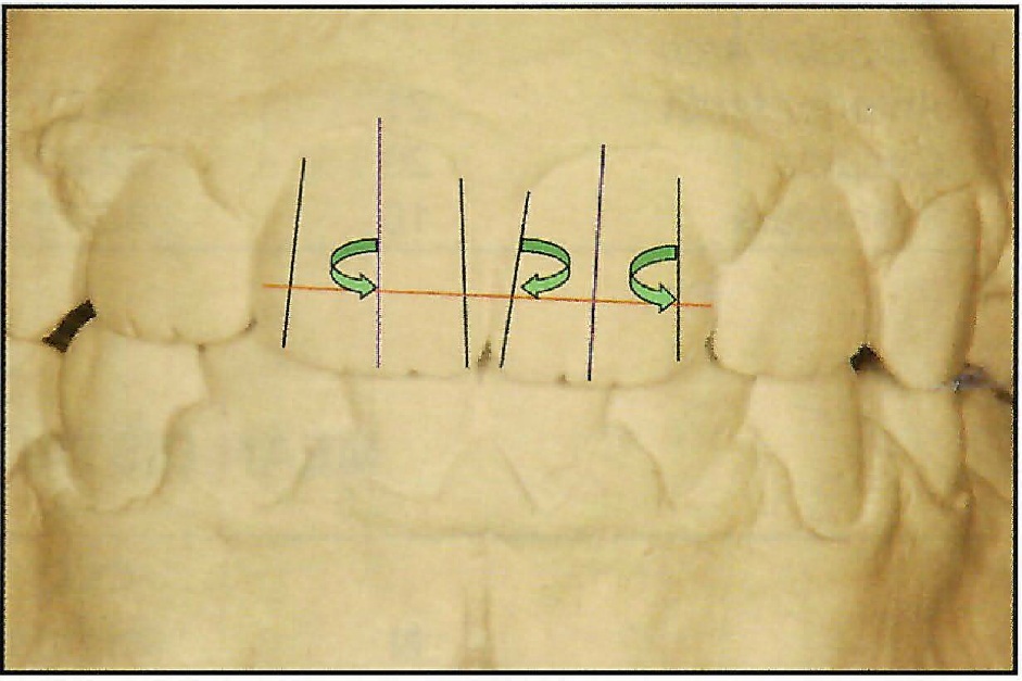
Fig. 3 Mesial and distal secants, crown long axis, and occlusal plane drawn on photograph of plaster cast.
Positioning Brackets with the Crown System
The mean secant angles, along with the normal anatomy of each tooth, helped us establish the contours of the bracket bases (Figs. 4 and 5).
In bonding to teeth with normal crown forms, the brackets are positioned so they are parallel to the imaginary mesial and distal secants (Fig. 6). With incisor brackets, the incisal edge, which is normally parallel to the occlusal plane, can be used as another reference. On the cuspid bracket, the tip of the bracket base points toward the cusp tip for further assistance in alignment.
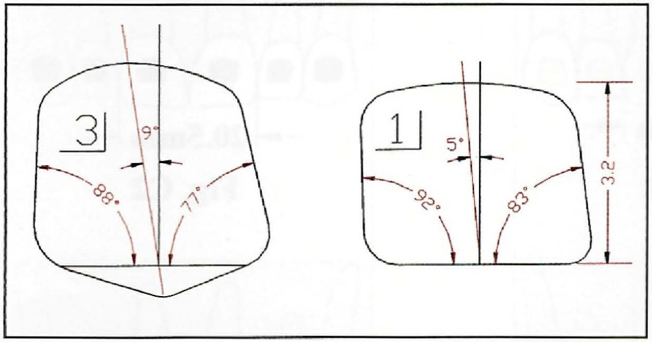
Fig. 4 Mean secant angles used to establish contours of maxillary right cuspid and right central incisor brackets.
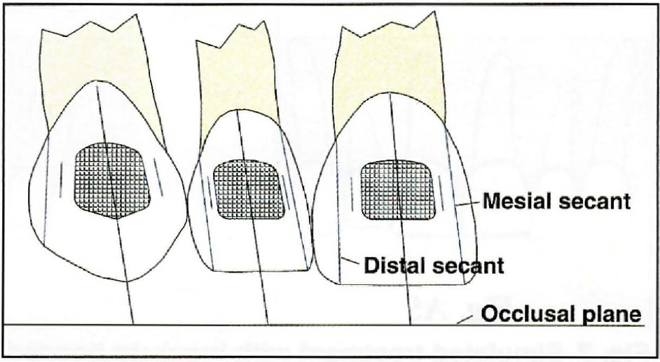
Fig. 6 Positioning brackets parallel to mesial and distal secants and incisal edges, which are normally parallel to occlusal plane. Cuspid bracket tip points toward cusp tip for aid in alignment.
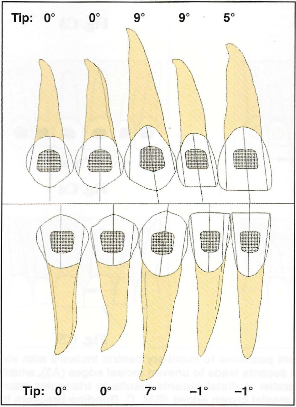
Fig. 5 Bracket contours shown true to scale.
The vertical bracket positions should correspond to the LA points of Andrews.3 Our usual procedure, however, is to start by placing the mandibular second bicuspid bracket as close as possible to the gingival margin. All other brackets are then bonded at the same distance from the incisal edges. The same approach is used in the maxillary arch.
Although human crown forms have common anatomical characteristics, they still display a great deal of individual variation. A major advantage of the Crown System is the early recognition of abnormal crown forms. If either secant or the incisal edge is not parallel to the contours of the bracket base, it is easy to detect which crown edge deviates from the norm or has been altered--for instance, by abrasion. This helps determine which edges of the bracket should be parallel and aligned and which edges of the crown should eventually be built up or recontoured. Thus, the system can avoid the need for rebonding while allowing the brackets to achieve their preprogrammed tip and optimum esthetics.
Through the early diagnosis of crown-form discrepancies, the clinician can use the bracket positions to influence the course of treatment and the final result. In the theoretical example shown of two maxillary central incisors with abnormal crown forms (Fig. 7), the brackets can be placed in three different positions, all of which will have different effects on crown and root angulation. Bonding the brackets parallel to the mesial secants, as in Row A, might be appropriate if the incisal edges had been ground down or damaged by trauma, or in case of a tooth-size discrepancy between the maxillary and mandibular arches. Recontouring a central incisor that was already too small, as shown in Row B, would produce an even greater size discrepancy. If the incisors were wide enough, however, the approach of Row B would be preferable. The Crown System helps the clinician make the right decision at the beginning of treatment.
Fig. 7 Simulated treatment with brackets bonded in different positions to maxillary central incisors with abnormal crown forms. A. Bonding brackets parallel to mesial secants leads to uneven incisal edges (A3), which requires composite build-up (A4). B. Bonding brackets parallel to distal secants results in triangular interdental space (B2), which can be resolved by recontouring mesial crown edges (B3). C. Bonding brackets in conventional centered positions creates both problems, even after recontouring (C3).
Case Report
A male patient age 9 years, 8 months, presented with a Class I malocclusion, crowding in both arches, a midline deviation, and the maxillary right first permanent molar in crossbite (Fig. 8).
Fig. 8 9-year-old male with Class I malocclusion before treatment.
Treatment was begun with 2 X 4 appliances in both arches, using .022" Crown Line brackets placed with the Crown Bonding System (Fig. 9). Total treatment time was 45 months (Fig. 10).
Fig. 9 Placement of .022" Crown Line brackets using Crown Bonding System, with central incisor brackets parallel to mesial secants.
Fig. 10 Patient three months after debonding.
REFERENCES
- 1. Kokich, V.: Esthetics and anterior tooth position: An orthodontic perspective, Part I: Crown length; Part II: Vertical position; Part III: Mediolateral relationships, J. Esth. Dent. 5:19-23, 174-178, 200-207, 1993.
- 2. Zachrisson, B.: Esthetic factors involved in anterior tooth display and the smile: Vertical dimension, J. Clin. Orthod. 32:432-445, 1998.
- 3. . Andrews, L.F.: The Straight-Wire Appliance, J. Clin. Orthod. 10:99-114, 174-195, 360-379, 1976.
-
 WOLFGANG HEISER, MD
 -
 CLAUS SCHENDELL, Dipl Ing
Dr. Heiser is in the private practice of orthodontics at Dr. Stumpf Str. 73, A-6020 Innsbruck, Austria. Both authors have a financial interest in the products mentioned in this article.
Mr. Schendell is President of Adenta GmbH, Gilching, Germany. Both authors have a financial interest in the products mentioned in this article.