THE EDITOR'S CORNER
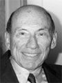
We have been fortunate in orthodontics to have had a large number of outstanding people attracted to our field. Among them is the gentleman who graces our cover this month in an office setting reminiscent of the time when he entered the practice of dentistry in 1919. He is, of course, Dr. Cecil Steiner, now of Laguna Hills, California. Dr. Steiner has been an important link between the founder of orthodontics, Dr. Edward Angle, with whom he studied, and the more recent generations of orthodontists, who studied with him. Those of you fortunate enough to know Dr. Steiner are well acquainted with those characteristics which caused him to stand out from the crowd.
At a time when orthodontics was developing its basic appliances, Dr. Steiner was extremely creative and inventive. His name is associated with improved edgewise bracket design, .018 bracket slot, Steiner Spring-Wing brackets, and numerous useful tools of our specialty. At a time when orthodontics was developing its basic science, Dr. Steiner developed the Steiner cephalometric analysis. At a time when orthodontics was developing its means of communication, Dr. Steiner was instrumental in the establishment of the Angle Society and the Tweed Foundation. He was unstinting in his travels and in his devotion to "spreading the word" at meetings, at study clubs, at graduate orthodontic departments here and abroad. It was this spirit of sharing which exemplified Dr. Steiner's contributions to the specialty he loves and which qualifies him for a place in the Orthodontic Hall of Fame.