Favorite Saved

Adult Orthodontics

In the last few years there has been considerable interest in orthodontic treatment for the adult patient. A recent survey conducted by the AAO showed an increase in the percentage of patients over 21 years of age from a fraction more than 4% ten years ago to almost 7% today. Nearly 11% is expected after another decade.

Since an adult is defined as one who is fully grown, most males of 18 or 19 and most females of 16 can be considered as adults. The percentage of adult patients would therefore be somewhat higher than the figures shown above. Over the past ten years in my own practice, 10% to 15 % of the patients have been adults.

Dr. Irving Glickman, the eminent periodontist, has often pointed out the need for orthodontic treatment for adult patients. As long as fifteen years ago, he made a strong plea to orthodontists attending the annual AAO meeting to accept adult patients on a routine basis.

What are the differences between adult and child orthodontics? The basic difference is that in children we must concern ourselves with tooth movement plus growth, whereas in adults we are dealing strictly with tooth movement. We cannot count upon growth to help us (or hinder us) in achieving our treatment objectives. In a way, then, adult orthodontics is simpler, for we have one less (and often unpredictable) factor with which to contend. There are, however, several other differences between adult and child orthodontics (Table I) .

Motivation

Why do adults seek orthodontic treatment? Based on my own practice experiences, I would list some of the reasons as follows:

  • Did not want orthodontic treatment as children.
  • Did not know about orthodontics as children.
  • Parents could not afford orthodontic treatment.
  • Were not advised by dentist of need for orthodontic treatment when- younger.
  • No orthodontist located in their vicinity when younger.
  • Incomplete orthodontic treatment as children. Non-cooperative.
  • Had orthodontic treatment as children, but relapsed.
  • More conscious of appearance with age.
  • Malpositioned teeth contributing to periodontal disease.
  • Increasing difficulties in mastication.
  • Malocclusion and mandibular slide producing soreness in the temporomandibular joint.
  • Spaces between anterior teeth enlarging, or new spaces opening up.
  • Anterior teeth starting to crowd or minor crowding becoming more severe.
  • For better tooth positioning prior to prosthetic restoration.
  • Prevention. Concerned about "keeping their teeth".
  • The recent periodontal awareness of the dental profession has indirectly affected the orthodontic specialty in that orthodontic treatment has now become an integral part of the total treatment plan concept. Therefore, close co-operation of the orthodontist with the periodontist and restorative dentist is essential for proper treatment of the adult patient.

    Procedure

    The usual sequence of procedure in adult patients is as follows:

  • 1. Eliminate all pathology (e.g. caries, abscesses, periodontal disease, retained roots, etc. ) .
  • 2. Orthodontic treatment.
  • 3. Periodontal re-evaluation (and therapy if necessary).
  • 4. Prosthetic restoration (when necessary) .
  • 5. Orthodontic retention (when necessary) .
  • 6. Periodontal maintenance.
  • Occlusal adjustments ( grinding) should be performed when ever necessary during all of the above stages.

    It should be pointed out that orthodontic treatment, whether it be for the adult or the child patient, is actually a type of mouth rehabilitation. When performed on the adult patient in conjunction with the restorative dentist, the orthodontic treatment is one aspect of the total oral rehabilitation for that patient. In some instances no restorative work is required following orthodontic treatment. Thus the total oral rehabilitation can be carried out orthodontically. In other instances the rehabilitation can be carried out solely by the prosthodontist (or restorative dentist) through the use of inlays, on-lays, crowns, bridges, splints, etc. Thus if the term "full mouth rehabilitation" applies to the prosthetic restoration of a mouth to proper health and function, it should apply equally to the orthodontic

    restoration of a mouth.

    Classification

    Since many of our adult patients require both orthodontic and prosthetic services, a classification has been devised to facilitate the treatment planning of adult patients (Table II) .

    CASES

    The following cases will serve as examples of the classification and illustrate the wide scope of adult orthodontic treatment.

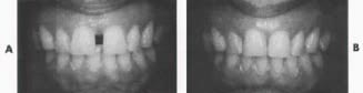
    Case 1 (Fig. 1) Mrs. P. S. Age 39

    CLASS A 1 (Minor orthodontics; orthodontic retention). Treatment time-- 7 months. Reason for treatment-- Wide diastema between upper centrals which was becoming a major concern to the patient.

    Orthodontic treatment--

  • 1. Moved upper right central mesially.
  • 2. Retracted six upper anteriors (3s only slightly).
  • 3. Moved upper 4s lingually.
  • 4. Ground labio-incisal of six lower anteriors and lingual of four upper incisors.
  • 5. Stripped six lower anteriors a bit.
  • 6. Equilibrated occlusion in centric relation, eliminating slight forward mandibular slide. Appliances--
  • Maxillary-- removable (high labial type). Mandibular-- none.

    Case 2 (Fig. 2) Mr.E.F Age 37

    CLASS B 3 (Minor orthodontic, prosthetic restoration; fixed splinting). Treatment time-- 5 months. Reason for treatment-- Dentist recommended orthodontic treatment for correction of crossbite prior to commencing prosthetic rehabilitation.

    Orthodontic treatment--

  • 1. Extracted upper right 8, upper centrals, upper left lateral and four lower incisors because of periodontal involvement.
  • 2. Contracted lower right 3,4,5,6,7. Appliances--
  • Maxillary-- removable with pontics replacing upper centrals and upper left lateral. Mandibular-- removable with pontics replacing four lower incisors and posterior bite plane to disarticulate teeth in crossbite.

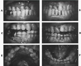
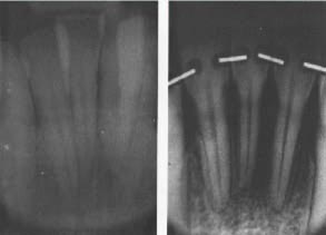
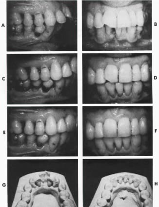
    Case 3 (Figs. 3 and 4) Mr. P. S. Age 38

    CLASS C 1 + 2 (Major orthodontics; orthodontic retention (maxilla), temporary splinting

    (mandible). Treatment time-- 22 months Reason for treatment-- Advice of periodontist. Severe lower anterior crowding contributing

    greatly to periodontal problem. Prognosis on four lower incisors uncertain. Better positioning of lower 3s advisable for future bridgework if lower incisors were to be lost.

    Orthodontic treatment--

  • 1. Lower right central extracted.
  • 2. Remaining lower anteriors aligned.
  • 3. Upper right 3,4,5,6,7 moved distally.
  • 4. Upper right 7 contracted.
  • 5. Upper incisors retracted.
  • 6. Stripped upper centrals. Appliances--
  • Maxillary-- removable (high labial type). Mandibular-- fixed bands.

    Case 4 (Fig. 5) Miss H. N. Age 30

    CLASS D 1 + 2 + 3 (Major orthodontics, prosthetic restoration; orthodontic retention and temporary splinting (maxilla), fixed splinting (mandible) . Treatment time-- 24 months. Reasons for treatment-- Patient concerned with overlapping upper central incisors. Dentist advised orthodontics for better tooth positioning prior to prosthetic restoration

    Orthodontic treatment--

  • 1. Upper left 1, 2, 3, 4, 5 moved distally.
  • 2. Upper left central rotated.
  • 3. Four upper incisors and lower centrals retracted.
  • 4. Lower 7s moved distally to upright.
  • 5. Overbite reduced.
  • 6. Upper right 7, lower left 8 extracted. Appliances--
  • Maxillary-- removable (with anterior bite plane). Mandibular-- removable (with posterior bite plane).

    Case 5 (Fig. 6) Mrs. M. C. Age 38

    CLASS B 1 + 3 (Minor orthodontics, prosthetic restoration; orthodontic retention, fixed splinting).

    Treatment time-- 8 months. Reason for treatment-- Severe posterior crossbite on the right side causing difficulties in mastication.

    Orthodontic treatment--

  • 1. Extracted lower right 8, lower left 4.
  • 2. Expanded lower right bicuspids and first molar.
  • 3. Contracted upper right bicuspids and first molar and upper left 4.
  • 4. Moved upper left 3 distally.
  • 5. Moved upper and lower centrals lingually.
  • 6. Moved lower right 2 labially. Appliances--
  • Maxillary-- removable. Mandibular-- removable.

    Case 6 (Fig. 7)Mr. H. H.Age 49

    CLASS C 1 (Major orthodontics; orthodontic retention) Treatment time-- 15 months.Reason for treatment-- Patient was advised by his dentist to have his anterior occlusion corrected orthodontically. Traumatic occlusion was causing occasional small fractures of maxillary incisors.

    Orthodontic treatment--

  • 1. Extracted lower right 3, lower left 4. Lower right 3 was chosen because of deep caries and possible pulp exposure.
  • 2. Moved lower left 3 distally and lingually.
  • 3. Moved lower right 2 lingually.
  • 4. Aligned lower anteriors torquing roots lingually as much as possible.
  • 5. Moved lower left 6 lingually.
  • 6. Ground linguo-incisal of upper centrals and upper left lateral for proper occlusion.
  • 7. Stripped lower anteriors slightly. Appliances--
  • Maxillary-- none. Mandibular-- fixed bands.

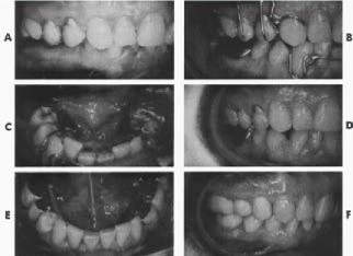
    Case 7 (Figs. 8 and 9)Mr. W. F.Age 45

    CLASS D 2 + 3 (Major orthodontics, prosthetic restoration; temporary splinting (anteriors), permanent splinting (posteriors) .Treatment time-- 17 months. Reason for treatment-- Recommended by dentist and periodontist for better tooth placement prior to prosthetic rehabilitation. Also for closure of diastema between upper centrals.

    Orthodontic treatment--

  • 1. Lower left 5 extracted.
  • 2. Lower left 4 moved distally and lingually.
  • 3. Lower 3s expanded a bit.
  • 4. Lower incisors retracted a bit.
  • 5. Upper left 4 moved buccally.
  • 6. Upper left 3, 5, 6, 7 moved lingually.
  • 7. Upper right 3, 4 moved distally.
  • 8. Upper incisors retracted.
  • 9. Upper left central rotated and uprighted.
  • 10. Bite opened a little on posteriors. Appliances--
  • Maxillary-- removable (high labial type). Mandibular-- removable (Hawley type) . Occupation of patient precluded use of fixed bands.

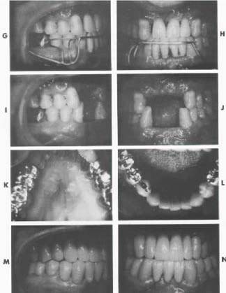
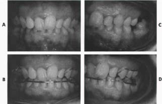
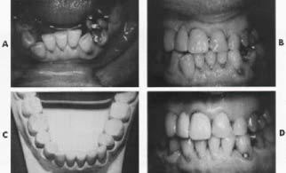
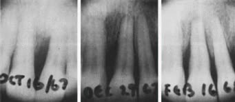
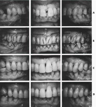
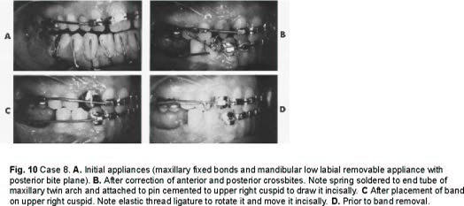
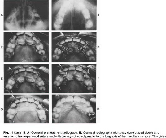
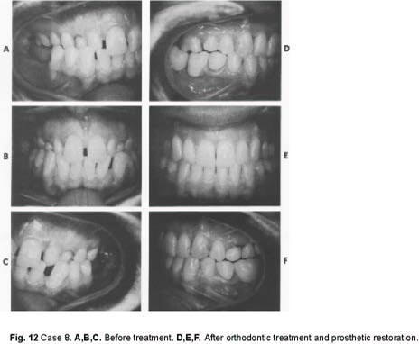
    Case 8 (Figs. 10, 11, and 12) Miss D. L. Age 26

    CLASS D 1 + 3 (Major orthodontics, prosthetic restoration; orthodontic retention, fixed splinting). Treatment time-- 33 months. Reason for treatment-- Patient concerned with the appearance of her teeth and the health of her mouth. She came from a small town where no orthodontic treatment was available. She now has an important position and wishes to do whatever is possible to improve herself. Orthodontic treatment--

  • 1. Upper right 4, 5, 6 and left 4, 5 expanded.
  • 2. Upper incisors aligned. Laterals moved labially.
  • 3. Lower 3, 4, 5s contracted.
  • 4. Lower incisors retracted and all six anteriors aligned.
  • 5. Upper and lower left bicuspids moved slightly mesially.
  • 6. Upper left 7 moved distally to upright.
  • 7. Upper Cs extracted, 3s surgically exposed, pinned, and moved orthodontically into alignment.
  • 8. Upper right 8 and retained roots in lower edentulous areas extracted. Appliances--
  • Maxillary-- fixed bands. Mandibular-- first removable, then fixed bands.

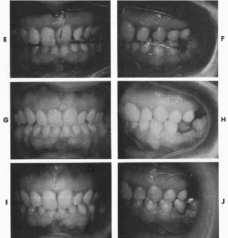
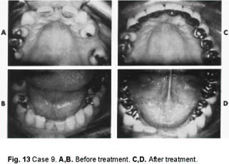
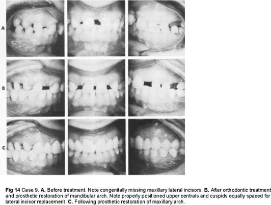
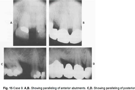
    Case 9 (Figs. 13), 14) and 15) Mrs. M. L. Age 38

    CLASS D 3 (Major orthodontics, prosthetic restoration; fixed splinting). Treatment time-- 17 months.Reason for treatment-- Patient was extremely self-conscious about the appearance of the anterior teeth, especially the upper central diastema which was due primarily to congenitally missing laterals. Upon accepting positions of responsibility in community organizations, she felt something had to be done about her teeth. Her dentist felt that proper results could not be achieved by bridgework alone.

    Orthodontic treatment--

  • 1. Upper 3s and upper left 4 moved distally.
  • 2. Upper centrals moved mesially.
  • 3. Upper centrals and cuspids, aligned, intruded and centered, leaving proper and equal space for lateral pontics.
  • 4. Lower six anteriors retracted slightly.
  • 5. Lower bicuspids moved mesially to close spaces.
  • 6. Lower 7, 8s moved distally to upright. Appliances--
  • Maxillary-- fixed bands.

    Mandibular-- removable.

    Conclusion

    There is a great need for orthdontic treatment for the adult patient. Continuing education of the general public will result in an increasing demand for this type of service. The orthodontist should update his knowledge and his thinking in this aspect of his responsibility, and should try as much as possible to co-ordinate his efforts with those of his confreres in other branches of dentistry in order to render to the population a better and more complete dental health service.

    Fig. 1 Case 1 A Before treatment B After treatment,
    Fig. 2a Case 2. A,B. Before treatment and before extractions. C,D. With appliances in place, before extractions. E,F. Occlusal views with appliances in place, before extractions.
    Fig. 2b Case 2 G,H. After correction of crossbite. Note pontics added to appliances. I,J. After orthodontic treatment, with appliances removed. K,L. Occlusal views after orthodontic treatment and prosthetic restoration. M,N. After orthodontic treatment and prosthetic restoration.
    Fig. 3 Case 3. A. Before treatment. B. After treatment. Note temporary splinting (A Splints), which may be replaced with permanent splints.
    Fig. 4 Case 3. A,B. Before treatment, after extraction of lower right central. C,D. After removal of mandibular fixed bands. E,F After orthodontic treatment, temporary splinting of lower anteriors, and restoration on upper right central. G,H. Lower anteriors before and after treatment.
    Fig. 5a Case 4. A,B. Before treatment. C,D. With appliances in place. Note posterior bite plane on lower appliance; springs to move upper left 3,4,5 distally; and buccal guide wire.
    Fig. 5b Case 4 E,F. After completion of distal movement of upper left 3,4,5. Note high labial wire and spring against mesiolabial of upper left central to rotate and retract it. Shortly after, hooks were soldered to high labial wire in cuspid regions and an elastic used for slight retraction of upper incisors. Lower appliance was also altered in order to upright lower 7s. G,H. After completion of orthodontic treatment. Note improvement in inclination of lower left 7. I,J. After prosthetic restoration. Upper centrals were temporarily splinted to aid in retention.
    Fig. 6 Case 5. A. Before treatment showing maxillary posterior crossbite. B. Appliances in place. C. Lower appliance before treatment. Note posterior bite platform. D. After orthodontic treatment. E, F. After prosthetic restoration.
    Fig. 7 Case 6. A,B. Before treatment. C,D. After treatment
    Fig. 8 Case 7. Periapical x-rays before uprighting (left) showing infrabony pocket and proximity of roots of left central and lateral; after two months (center) showing improvement; and after four months (right) left central is uprighted and roots of left central and lateral are separated.
    Fig. 9 Case 7. A. before treatment. B. With upper and lower removable appliances in place. Note posterior bite plane on lower appliance to restore lost vertical dimension. C. After orthodontic treatment, periodontic treatment, and temporary stabilization. Elimination of deep pocket has resulted in lengthening of clinical crown of upper left central. D. Completed case after prosthetic restoration.
    Fig. 10 Case 8. A. Initial appliances (maxillary fixed bonds and mandibular low labial removable appliance with posterior bite plane). B. After correction of anterior and posterior crossbites. Note spring soldered to end tube of maxillary twin arch and attached to pin cemented to upper right cuspid to draw it incisally. C After placement of band on upper right cuspid. Note elastic thread ligature to rotate it and move it incisally. D. Prior to band removal.
    Fig. 11 Case 11. A. Occlusal pretreatment radiograph. B. Occlusal radiography with x-ray cone placed above and anterior to fronto-pariental suture and with the rays directed parallel to the long axis of the maxillary incisors. This gives o true cross-sectional appearance and more accurately locates impacted canines. C. Initial appliances. D. Following exposure and pinning of upper cuspids. Note elastic thread ligature from pins to twin wire. E. Cuspids approaching alignment. F. Bands cemented to cuspids. Elastic thread ligature used for rotation. G. Cuspid alignment almost completed. Note acrylic button added to Mershon lingual appliance for additional anchorage. H. Completed maxillary arch.
    Fig. 12 Case 8. A,B,C. Before treatment. D,E,F. After orthodontic treatment and prosthetic restoration.
    Fig. 13 Case 9. A,B. Before treatment. C,D. After treatment.
    Fig. 14 Case 9. A. Before treatment. Note congenitally missing maxillary lateral incisors. B. After orthodontic treatment and prosthetic restoration of mandibular arch. Note properly positioned upper centrals and cuspids equally spaced for lateral incisor replacement. C. Following prosthetic restoration of maxillary arch.
    Fig. 15 Case 9. A,B. Showing paralleling of anterior abutments. C,D. Showing paralleling of posterior

    DR. HARVEY L. LEVITT DDS, FRCD (C)

    DR. HARVEY L.  LEVITT DDS, FRCD (C)

    My Account

    This is currently not available. Please check back later.

    Please contact heather@jco-online.com for any changes to your account.