Favorite Saved

Bonding Attachments Directly to Teeth

The placement of orthodontic attachments (buttons, hooks, and other attachments) directly to the teeth without the necessity of fabricating bands has long been the orthodontist's dream. For the past ten years, the Orthodontic Department at the Bronx-Lebanon Hospital Center has successfully developed and utilized the technique described in this article. It has been used most effectively in the anterior part of the mouth where esthetics is so important, or in specific situations where banding is very difficult or impossible. To date, the adhesive properties of a specific cold-cure acrylic called Pearl-on has been found to be most practical.

Indications

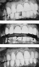
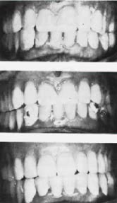
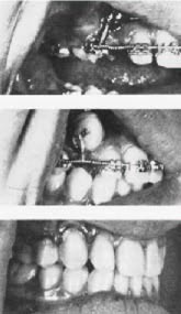
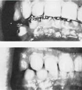
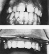
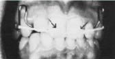
1-- In adult orthodontics preliminary to mouth reconstruction, especially the closing of anterior spaces and improving the axial inclination of the incisors (Figs. 1 & 2).2-- Where teeth cannot be banded easily:

  • a. Abutment teeth on a fixed bridge.
  • b. Where there is short crown height due to incomplete eruption or only partial surgical exposure (Fig. 3).
  • c. On malposed impacted teeth where the surgeon cannot place a ligature tie-wire around the cervical margin (Figs. 4 & 5) .
  • d. On partially erupted or surgically exposed posterior teeth (Fig. 6) .
  • e. In cases where surgical tie-wire has broken (Fig. 5) and when the impacted tooth still has insufficient crown length (Fig. 7) .
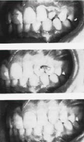
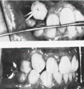
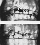
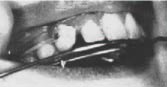
  • 3-- For intrusion, elongation and retrusion of anterior teeth while wearing the face bow appliance; in situations where it is desirable to avoid anterior bands and still maintain adequate control of tooth movement (Figs. 8, 9 & 10).

    Technique

  • 1. The desired attachment is selected (metal buttons, weldable Unitek molar hooks, twisted brass ligature wire hooks, flanged molar buccal tubes, or plastic brackets).
  • 2. The individual tooth designated to carry the attachment is thoroughly cleaned with a fluoride tooth paste, air-dried, and the labial surface is covered with a drop of regular cement liquid (orthophosphoric acid) for 45 seconds. This produces a microscopic etching on the tooth surface.
  • 3. The area is then washed with a stream of water and air dried again. The area must be well isolated with cotton rolls and kept dry from seepage of saliva or blood.
  • 4. Pearl-on acrylic (shade 21) is mixed in a dappen dish with the monomer liquid and immediately
  • applied to the tooth surface with a fine camel-hair brush. After the first thin layer has been exposed to the air for a few seconds, an additional amount of acrylic mix is placed on the same spot.

  • 5. The attachment selected is then embedded into the acrylic, using college pliers, covering the base only. If necessary, add more acrylic to cover the entire base.
  • 6. In about five minutes the excess acrylic may be polished away with a large white burlew wheel and generally smoothed.
  • 7. To insure maximum retention of the Pearl-on, pressure should not be applied until the following visit.
  • After treatment is accomplished or normal band placement is made possible, the acrylic base and attachment may be removed by means of a sharp scaler or with an ultrasonic prophylaxis instrument such as Cavitron. No damage has been known to take place on the enamel surface.

    Commercially produced plastic brackets have been success fully attached using Pearl-on acrylic as the bonding medium and the attachment has been strong enough to hold light wires, up to .016", for periods up to six months and longer.

    Experimental work is now being conducted with an alternate product called Permite A, shade 65. Both products are available through any dental supply house and are manufactured by American Consolidated Manufacturing Co., Inc., Philadelphia, Pa.

    Summary and conclusions

    By means of the technique described, various attachments may be successfully bonded to teeth by means of Pearl-on cold cure acrylic. The improved esthetics and the simplicity of the procedure make it a useful adjunct in both general and specialty practice. It is a boon to orthodontists in situations where it is not practical to place bands.

    Fig. 1 (Above} Drifting of teeth in a 50-years old patient presents a problem in fabricating a lower six-unit splint. (Middle) Molar hooks were attached directly to the lower cuspids and to the lower right central and lateral incisors with Pearlon. Using elastics on the cuspids and elastic thread ligature on the incisors, in a few visits the spaces were evenly distributed and the teeth prepared for jacket crowns, (Below) The finished six-unit splint in place.
    Fig. 2 (Above) A 55-yeor-old patient with a full upper denture and a partial lower with drifting lower anterior teeth due to bone loss. (Middle) Buttons were attached directly to the cuspids with Pearl-on and an elastic was used to close spaces and upright the lower anteriors. (Below) tower anterior splint in place with new precision partial denture and new upper full denture.
    Fig. 3 (Above) A 16-year-old patient with a partially erupted, rotated cuspid. (Middle) A rotating spring was attached to the first bicuspid and a molar hook to the distal of the cuspid. (Below) The cuspid brought into alignment and rotated
    Fig. 4 (Above) Molar hook embedded in Pearlon covering labial surface of locked-out cuspid. A light ( 'h") elastic is extended from this hook to the molar bond. Extraoral appliance is used to reinforce anchorage. (Below} Cuspid in position immediately after band removal.
    Fig. 5 (Above) Closed coil. (.008x .036) tied from main archwire to cervical tie on impacted cuspid, (Middle) Cervical tie wire broke and was replaced with molar hook embedded in Pearl-on. Ligature wire is tied from hook to main archwire. {Below) Cuspid in position prior to cervical correction of gingival contour.
    Fig. 6 (Above) Unitek molar hook attached with Pearl-on to surgically exposed lower right first bicuspid, Note deflection of archwire to indicate degree of pressure that Pearl-on attachment will tolerate. (Below) Completed repositioning of the lower right first bicuspid,
    Fig. 7 (Above) Hook attached to labial surface of surgically exposed central incisor and tied to main archwire with elastic thread ligature. ( Below) The central incisor now tied directly to the main archwire and ready for banding.
    Fig. 8 Brass hook attached to cuspid with Pearl-on. Cuspid is moved distally with a latex elastic from the hook to the distal end of the extraoral appliance.
    Fig. 9 (Above) Pearl-on placed on upper centrals for better control of anterior latex elastic used on extraoral appliance. (Below) Space closure completed. Pearl-on has prevented elastic from riding into gingival tissue
    Fig. 10 Pearl-an buttons on upper central and upper left lateral for intrusion and retrusion with anterior latex elastics on a modified Hawley appliance.

    DR. IRVING GRENADIER DDS

    DR. IRVING  GRENADIER DDS
    Chairman and Attending, Orthodontic Department

    DR. CHRIS PHILIP DDS

    DR. CHRIS  PHILIP  DDS
    Associate Attending, Orthodontic Department

    DR. SAMUEL H. STEIN DDS

    DR. SAMUEL H.  STEIN DDS
    Associate Attending, Orthodontic Department

    My Account

    This is currently not available. Please check back later.

    Please contact heather@jco-online.com for any changes to your account.